Ambispora leptoticha (acaulosporoid morph)
Whole Spores
| Acaulosporoid morph | |
|---|---|
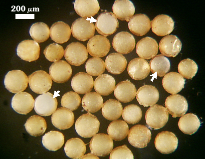 | 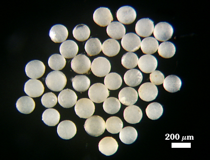 |
 COLOR: Cream (0-0-15-0) to dark orange-brown (0-40-100-10), most orange-brown (0-30-80-0), when all spore wall layers are present; bright white when outer two layers of spore wall have sloughed
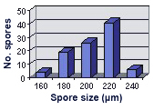
SHAPE: Globose, subglobose.
SIZE DISTRIBUTION: 160-240 µm, mean = 205.4 µm (n = 96)
Subcellular Structure of Spores
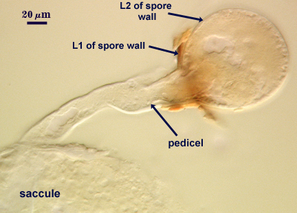
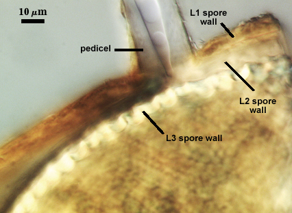
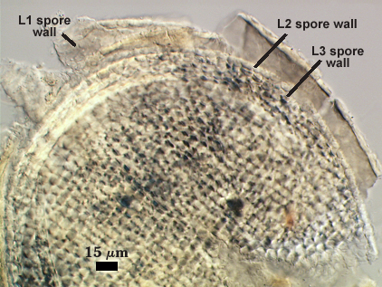
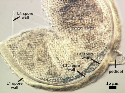
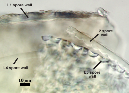
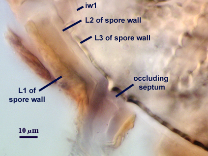
SPORE WALL: Four layers (L1, L2, L3 and L4), synthesized somewhat out of sequence (see photos below). L2 forms first, and it is just an expansion of the wall of the parent hypha, or “pedicel”. The outermost layer, L1, then is synthesized on the spore and slightly down the length of the pedicel. Time of synthesis is easily pinpointed by sudden appearance of red coloration caused by reaction of L1 in Melzer’s reagent. L3 is formed as a true endospore because it has no pore or connection to parent spore structures and seals off the spore contents. L4 is the last to form, and it is semi-flexible and has no pore or other discontinuity.
| Leptoticha morph | ||
|---|---|---|
|
|
|
| Leptoticha morph in Melzer's Reagent | ||
|---|---|---|
|
|
|
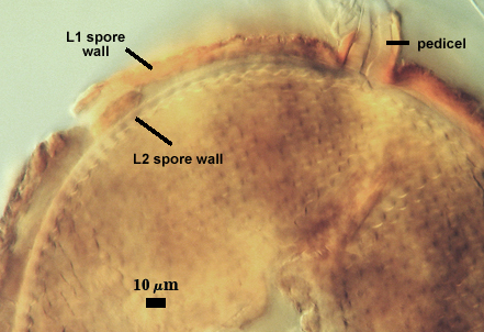
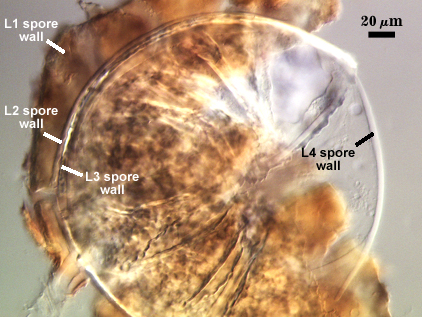
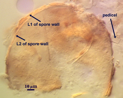
L1: A friable mass with deep fissures in the surface that breaks apart easily with applied pressure, expanding somewhat in PVLG-based mountants; hyaline in youth, becoming pale orange-brown (0-20-40-0) with age; 5.6-10.2 µm thick when intact, up to 14 µm in an expanded state; degrading and/or sloughing with age; staining dark orange-brown (0-60-100-10) to red-brown (20-60-100-0) in Melzer’s reagent. This layer forms after a spore has begun to develop from the pedicel and extends only a short distance on the “pedicel” (see developmental sequence above).
L2: Hyaline, semi-rigid, continuous with the wall of the pedicel; 3.8-5.6 µm thick; forming rounded convex hemispherical protruberances on the inner surface, 0.8-1.4 µm high that interlock with the concave depressions in L3 (below). This layer is continuous with the wall of the pedicel. While it does not degrade or break apart (like the outer layer), it still has a tendency to slough completely, leaving a shiny white endospore.
L3: Hyaline, rigid, consisting solely of tightly packed concave hemispherical depressions 3.2-6.2 µm wide and 2.9-4.6 µm deep (width of the layer). It forms just like the laminate layer in all sessile spores of Acaulospora species in that it is forms without any connection to previous layers (L1 and L2) and has no pore (thus no cytoplasmic link to the parent hypha). This layer is all that remains after L1 and L2 have sloughed; spores then are sessile, bright white and shiny, 160-200 µm in diameter (mean of 186 µm, n = 56).
INNER WALL Hyaline, semi-flexible, 3.8-5.0 µm thick (mean = 4.4 µm); consisting of multiple sublayers that often split into an outer thin layer, a thicker middle layer, and a thin inner layer; no reaction in Melzer’s reagent. This layer is labelled as L4 of the spore wall in some photos, and this is an incorrect interpretation. This error will be corrected soon.
| Spores mounted in PVLG | ||
|---|---|---|
|
|
|
| Spores mounted in Melzer’s reagent | ||
|---|---|---|
|
|
|
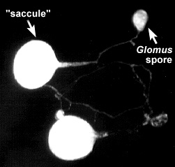
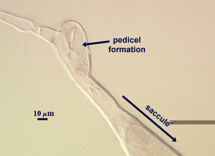
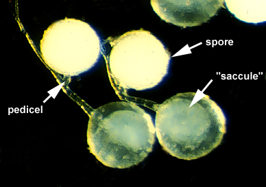
Pedicel
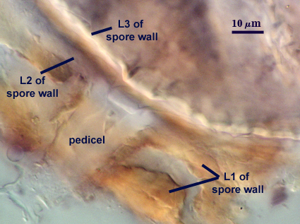
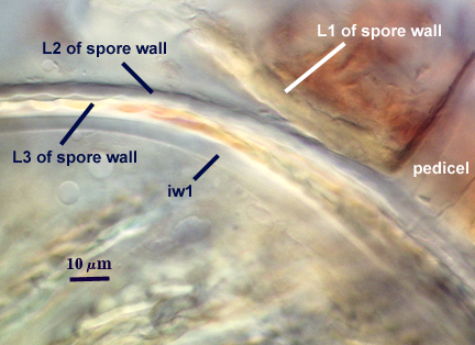
PEDICEL: So named because it is a feature divergent from most Acaulospora species, arising as a branch from the neck of the “sporiferous saccule”, 180-240 µm from the “saccule”. The pedicel is 38-64 µm in length, 22.6-32.4 µm wide at the base of the spore. Just prior to formation a distal thin septum forms that closes off contents of the pedicel and “saccule” from parent hyphae.
PEDICEL WALL: Two hyaline layers, with the outer layer (L1) continous with L1 of the spore wall and sloughing and the inner layer (L2) permanent and continuous with L2 of the spore wall. L1 is 4.5-7.2 µm thick at the base of the spore, tapering to < 2.0 µm at the juncture of the saccule neck. L2 is 4.2-6.3 µm thick at the base of the spore.
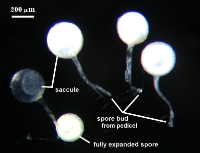
Sporiferous Saccule
COLOR: White (in youth) to hyaline (at maturity).
SHAPE: Globose to subglobose.
SIZE DISTRIBUTION: 240-320 µm, mean = 284.6 µm (n = 40)
SACCULE WALL: Hyaline and of variable thickness due to patchy sloughing of material so that the outer surface appears flaky; 4.0-6.8 µm thick.
DISTANCE FROM SACCULE TO SPORE: 180-240 µm.
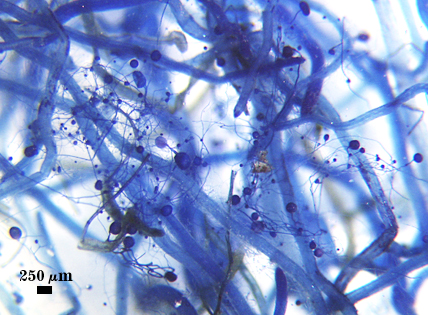
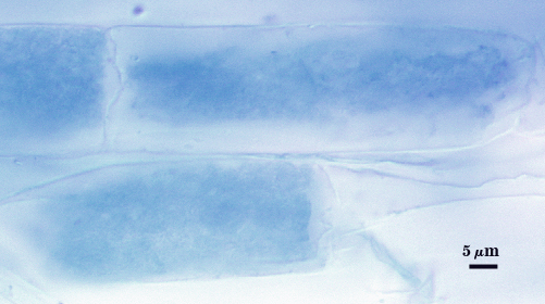
Mycorrhizae
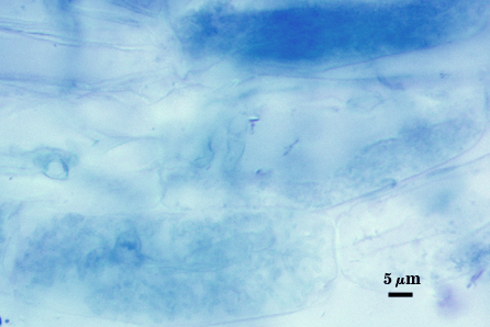
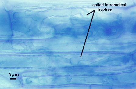
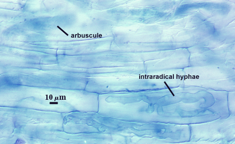
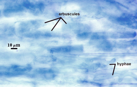
Coiled and knobby hyphae, 3.5-8.0 µm in diameter, with finely branced arbuscules in cortical cells. No vesicles have been observed in the four accessions of INVAM examined to date. The staining reaction of fungal structures in Melzer’s reagent is highly variable, from invisible to a very faint pale blue that often is hard to detect under a stereomicroscope (mounted roots are better). Contrast and color in the photos below were digitally enhanced to view structural details that often are hard to see clearly.
| Arbuscules in cortical cells of corn roots | ||
|---|---|---|
|
|
|
| All mycorrhizal structures in corn | |
|---|---|
|
|
Notes
Isolates of this species are prolific sporulators, with the occurrence of either or both synanamorphs being highly unpredictable. Some cultured isolates consistently produce one or the other morphotype; others produce both in varying proportions. Only occasionally are we able to mount a hypha bearing both synanamorphs (see photos).
| Prolific sporulators | |
|---|---|
|
|