Spore Germination
In the 1980’s, spore germination was examined in a wide range of mycorrhizal fungi. Methods included germinating spores on agar, on membranes above soil or soil solutions or in soils, and in root organ cultures. We study spore germination either for qualitative taxonomic analysis (to observe mode of germination) or to quantitatively examine tolerance of spores to varying culture conditions.
| Mycorrhizal fungi | |
|---|---|
 | 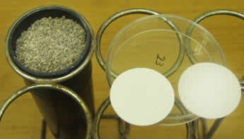 |
Spores are extracted, washed repeatedly in tap water, and agitated in a beadbeater for 4-5 minutes at 4800 rpm if there is considerable surface debris. A membrane filter (0.45 µm pores) is premoistened, 25-30 spores are collected in a pasteur pipette and transferred to the filter under a stereomicroscope.
Spores are gently redistributed, if necessary, so that none are touching (left photo above).
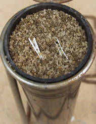
A deepot is filled with a presterilized sand and soil mix (4:1 v/v) (right photo above).
| Filled deepot | ||
|---|---|---|
 | 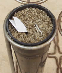 | 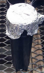 |
Filters are folded in half and then in half again (with the spores inside). They then are buried in the sand-soil mix of the deepot, moistened with sterile distilled water, covered with foil, and then placed in a rack in greenhouse or growth room.
Filters are gently removed after 2-3 weeks, opened, and placed in a glass petri dish containing hot 0.05% direct blue stain. After immersion for 30 seconds, the filter is transferred to a clean petri dish and examined.
Some spores with intact germ tubes are manually picked up with fine-tipped forceps and transferred to glass slides for permanent mounts in PVLG.
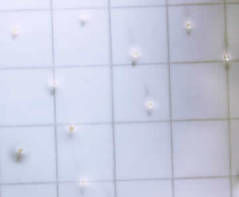
Photos of germinated spores (most with extensive hyphal development due to longer incubation times).