Mean Infection Percentage (MIP) Method
This assay (Moorman and Reeves, 1979) measures the percentage mycorrhizal colonization in an assay host after a limited time period so that only primary infections occur. Sometimes, assays must be conducted at different sample dilutions because the higher the infectivity, the sooner secondary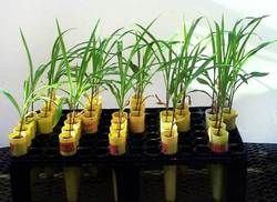 colonization occurs (which no longer separates infectivity from other host variables such as compatibility).
The MIP is a comparative assay in which a set of assays are measured relative to a reference assay (usually some sort of positive control such as a culture known to have good fungal growth). There is not a 1:1 correspondence between number of infectious propagules and the assay result. For example, if one wishes to compare commercial inoculants (all of which differ in important variables such as fungal species composition, properties of isolates in this species mix, and substrate composition), then a reference assay would be an inoculum of known properties.
In each assay, the statistic being measured can be either percentage colonization or mycorrhizal root length. The method of measuring % colonization by counting number of mycorrhizal segments out of 20 taken from a subsample of total roots per growth tube is not very accurate at lower dilutions. The preferred method for lower dilutions, is the grid-line intersect method (Giovannetti and Mosse, 1980) using a weighed subsample of roots. Mycorrhizal root length is a required metric if seedlings in each assay differs in host or growth medium because root biomass could differ greatly enough from one assay to the other that root length must be taken into account.
This assay has the advantage over the MPN of requiring fewer tubes to obtain a result, but it is equally sensitive to environment and the other host-soil variables. The trade-off is the more labor-intensive task of carefully estimating percentage of mycorrhizal roots in each tube of the MIP rather than scoring for presence/absence across all tubes of a replicate in the MPN.
Our MIP Assays are Conducted as Follows:
- Corn or Sudan grass are our standardized assay hosts because it is highly mycotrophic, produces abundant roots in three weeks, and fungal structures in the roots are easy to stain and visualize. If we are planning an experiment with a specific host species and we want to equalize inoculum density in that experiment, we use the experimental host in the assay.
- Plants are grown in plastic cone-tainers (4×18 cm), four replicates per assay. Usually 2-3 additional replicates are set up to examine % colonization at 30 days to determine if the assay should be harvested at that time or maintained for another 30 days (30 days is a maximum because of complications from secondary colonization, especially if different fungi are present amongst assays).
- Diluent consists of sterile sand (if changes in medium chemistry are important) or a mix of sterile sand and low-P, low organic matter soil (2:1 v/v) adjusted to pH 6.2. If the goal is to determine infectivity of two or more fungi to be used in an experiment, then the growth medium chosen for the experiment should be used.
- When samples are assumed to be highly infective, two dilutions generally can be used: 1/10 and 1/20. In our experience with testing field soils, pot culture inocula, and commercial inoculants, a 1/10 dilution is adequate. It is important to set up the assays so that ALL relevant sample comparisons are conducted at the same time. With only one dilution, this creates a better design to maximize number of treatments.
- Assays are carried out in a growth room with controlled light, temperature, and humidity conditions
- Roots are harvested, stained, and mycorrhizal colonization measured at either 21 days or 30 days. On occasion (mostly commercial inoculants obtained from users rather than a company representative), infectivity is so low that we extend the assay for 45 days to see if any propagules in the medium are active. One way to be sure of the target harvest date is to set up 2-3 extra tubes for each assay, and harvest these over time to pinpoint when secondary spread (merging of infection units) begins to appear.
- Usually, we measure percentage colonization using the grid-line intersect method in plastic petri dishes, because samples being compared for infectivity do not cause differential growth responses in the assay host. In cases where treatment samples being compared differ greatly in background chemical (and sometimes physical) properties, we measure mycorrhizal root length.
In the above MIP assay, six different inoculant formulations are being tested (with a nonamended control on the far left. The stimulatory effects of amendments are quite obvious in the middle three tubes. The two formulations on the far right are having a severe inhibitory effect on plant shoot and root growth and assay results invariably are negative (plants usually die and the assay terminated). If the fungi present in all of these treatments grew at a similar rate (if the formulation ingredients didn’t have a similar effect on the fungi, which were the same in all samples except the control), then percentage colonization would be lower in cones with high root biomass and higher in cones with fewer roots. To get a good measure of the proportion of roots colonized, fungal colonization must be compared against total root length.
References
- Moorman, T. and F. B. Reeves. 1979. The role of endomycorrhizae in revegetation practices in the semi-arid West. II. A bioassay to determine the effect of land disturbance on endomycorrhizal populations. Amer. J. Bot. 66: 14-18.
- Giovannetti, M. and B. Mosse. 1980. An evaluation of techniques for measuring vesicular arbuscular mycorrhizal infection in roots. New Phytol. 84: 489-500.