Glomus ambisporum
Voucher Specimens
This species is known in the culture collection only from type materials examined at Oregon State University. The photographs below were taken of these specimens. The description is taken from the protologue (Smith and Schenck, 1985) with some modifications to accommodate terminology defining development-based characters.
Whole Spores
Sporocarps dark brown to black, subglobosc to highly variable in shape, 315-690×424-776 µm, consisting of a single layer of spores originating from a central core of thick interwoven hyphae; peridium absent. Sporocarp aggregates up to 18×12 x 2 mm in size. Some sporocarps originating from and connected by a broad (20 µm) thick-walled (3 µm) hypha.
Spores with two distinct phenotypes are produced, both of which are glomoid in mode of formation and structure.
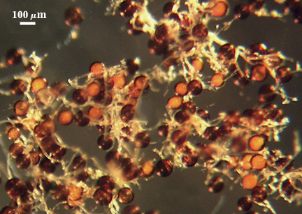
(1) Spores produced solely in sporocarps or aggregated around roots aredark brown to black, predominantly globose, 85-157 µm diam, to occasionally subglobose, 98-166×93-157 µm; maturing asynchronously budding from a sporogenous hypha.
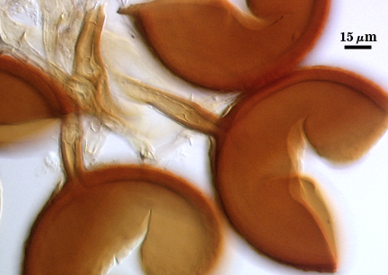
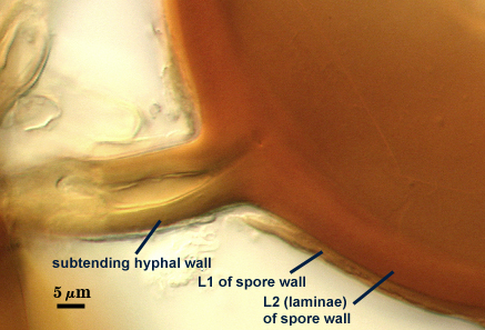
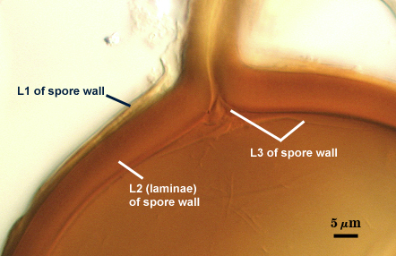
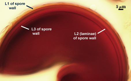
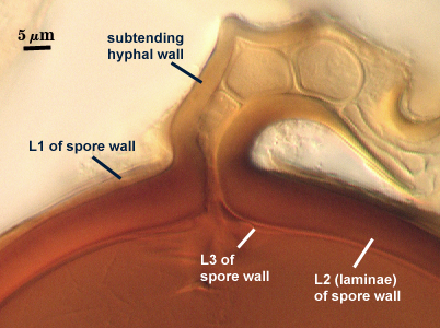
The spore wall consists of three layers. The outer layer (L1) is subyaline, 2-4 µm thick, extending the entire length of’ the hyphal attachment to the center of sporocarp. It has a reticulate surface described as consisting of an ordered arrangement of hexagonal plates 3-9 µm wide; often fracturing near the point of hyphal attachment oil detached spores leaving behind a collar circumscribing the attachment. The middle layer (L2) consists of finely adherent sublayers (or lamineae), 3-14 µm thick, dark brown to black in color, confluent with hyphal attachment. The innermost layer (L3) is thin and flexible (< 1 µm thick); originated from the innermost sublayer of the subtending hyphal wall (a continuation of L2 of the spore wall).
Subtending Hypha
The subtending hypha at the spore base is 10-24 µm wide; the wall consisting of two layers continuous with the outer two layers (L1 and L2) of the spore wall.
The occlusion consists of thickening of L2 together with L3 of the spore wall.
| In PVLG | In Melzer's | |||
|---|---|---|---|---|
 |  |  |  |  |
The spore wall consists of two layers, neither of which are reactive in Melzer’s reagent. hyaline to subliyaline, 2-4 pill thick. The outer layer (L1) is hyaline to subhyaline, consisting of fine sublayers (or laminae) up to 4 µm thick, breaking down and sloughing with age. The inner layer (L2) is hyaline to subhyaline, semi-flexible and 0.4-1 µm thick (the permanent layer?). Judging by the structure of attached hyphae in the voucher specimen, this layer appears to continue as part of the hyphal wall (often separating from the outer layer).(2) Spores formed singly or in unordered sporocarps in roots or in dead plant tissue. None were available in the type material to study. They are described as being subhyaline to hyaline, globose to subglobose, ellipsoid or highly variable in shape, 54-197×44-163 µm diam.
The subtending hypha at point of attachment 5-10 µm wide, consisting of two wall layers that are a continuation of the spore wall.
| Hyphae in the voucher, possibly associated with the unpigmented spore morphtype | |
|---|---|
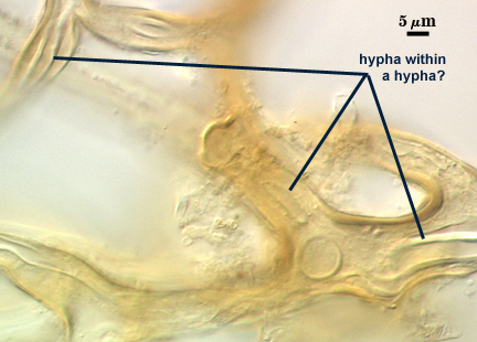 | 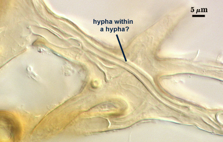 |
Reference
- Smith, G. S. and N. C. Schenck. 1985. Two new dimorphic species in the Endogonaceae: Glomus ambisporum and Glomus heterosporum. Mycologia 77:566-574.