Racocetra castanea
(reference accession BEG1)
Whole Spores
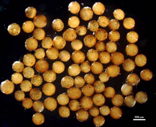
COLOR: Pale to dark copper (0-20-40-0) to slighly darker cream color (20-60-70-10).
SHAPE: Globose to subglobose.
SIZE DISTRIBUTION: 240-350 µm, mean = 320 µm (n = 100).
Subcellular Structure of Spores
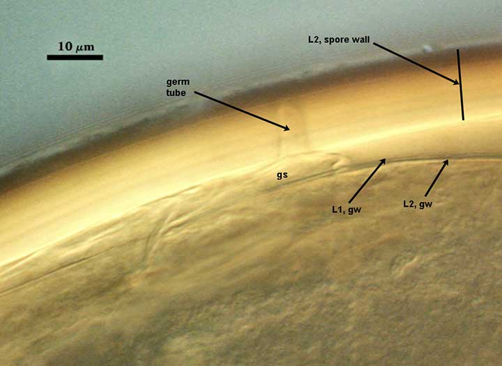
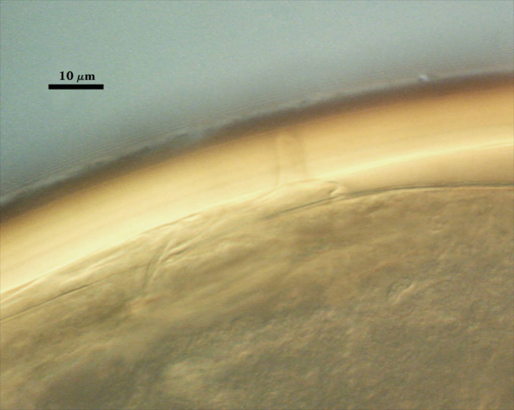
SPORE WALL: Two adherent layers (L1 and L2), with the inner laminate layer thicker than the outer layer.
| Spores mounted in Melzer’s reagent | |
|---|---|
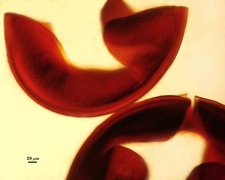 |  |
L1: An outer permanent rigid layer, yellow-brown (0-20-80-0), 0.7-1.8 µm thick, with a smooth surface.
L2: A layer consisting of orange-brown (0-30-80-0) to dark orange-brown (0-60-100-0) sublayers (or laminae) that increase in number with thickness, 6-9 µm thick (mean of 8 µm) in mature spores. This layer stains an orange-red (0-60-80-0) to red-brown color (20-80-80-0) in Melzer’s reagent.
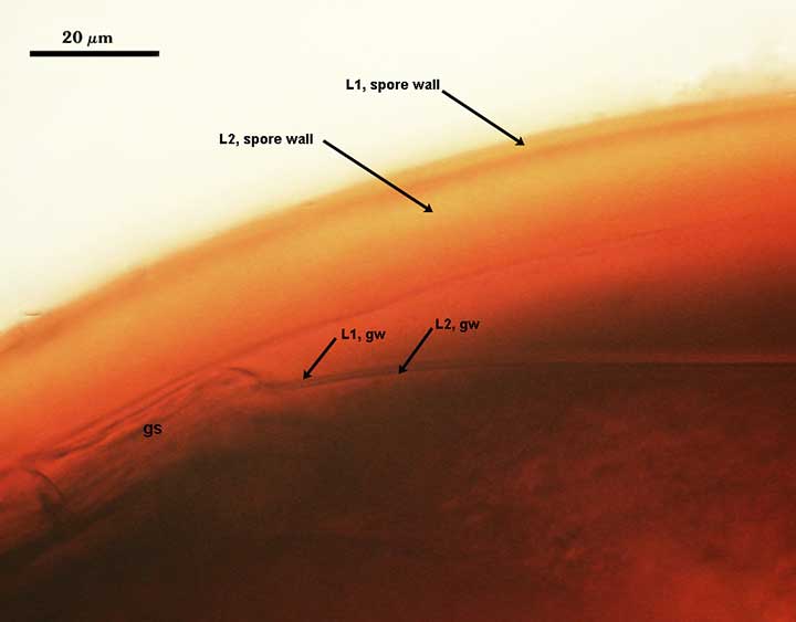
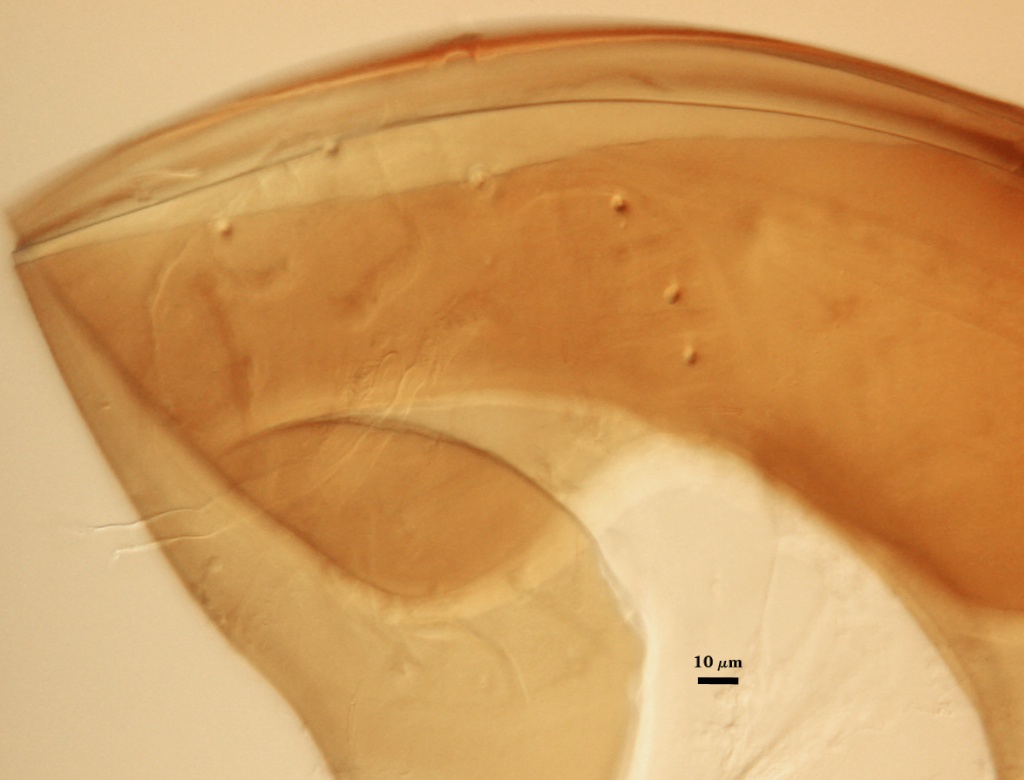
GERMINAL WALLS: One that is bi-layered, flexible, forming separately after the spore wall has fully differentiated. The germination shield develops on the surface of this wall.
| GERMINAL WALLS | |
|---|---|
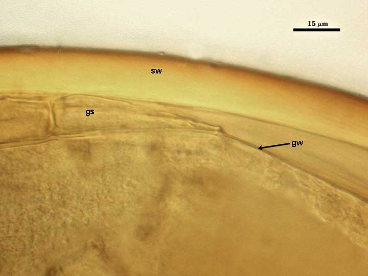 |  |
GW1: Two thin layers (L1 and L2) that usually are tightly adherent that they can appear as just one unit structure. Both layers usually can be detected in the region of the germination shield if it has formed. L1: A very thin layer less than 0.5 µm thick and thus difficult to resolve without differential interference optics. L2: A slightly thicker layer, 0.6-1.5 µm thick. Neither layer reacts in Melzer’s reagent.
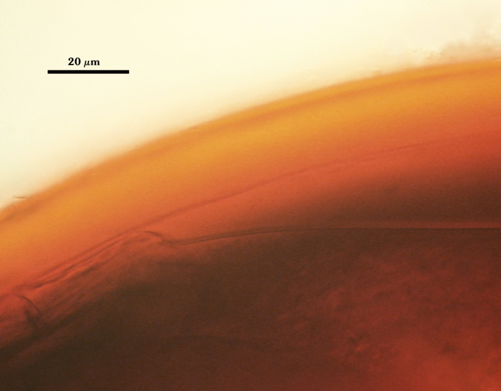
Subtending Hypha
WIDTH OF SPOROGENOUS CELL: 35-45 µm (mean = 40 µm).
SPOROGENOUS CELL WALL: Two hyaline layers (L1 and L2) probably are present (continuous with the two layers of the spore wall), but only L2 is readily discernible at the level of the compound microscope.
L2: Orange-brown (0-30-100-0), 2.4-5 µm thick near the spore and then thinning to about 1.5 µm beyond the sporogenous cell.
OCCLUSION: Closure by a plug concolorous with the laminate layer of the spore wall.
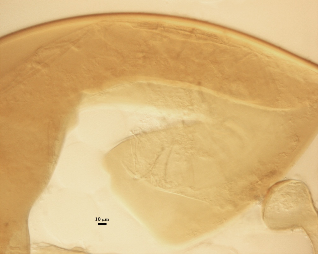
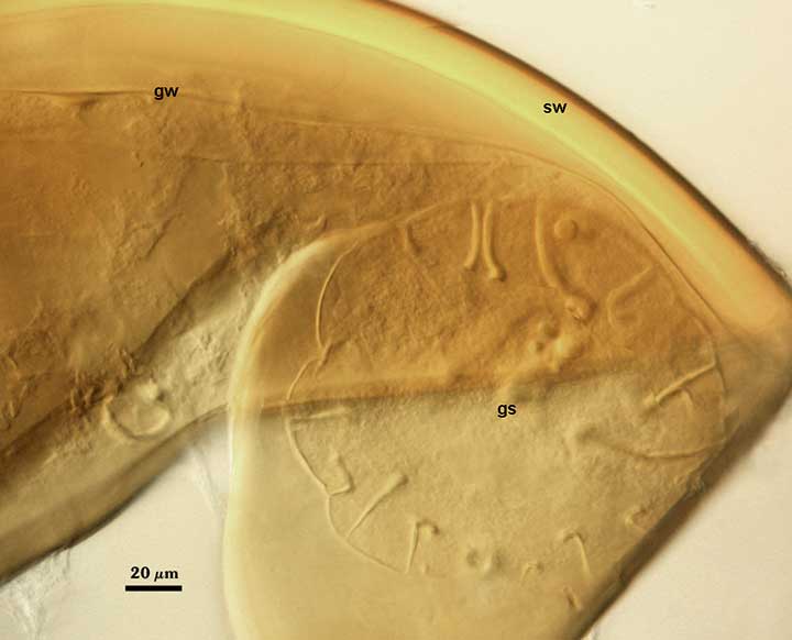
Germination
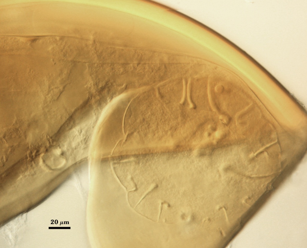
COLOR: Hyaline to pale yellow (0-0-20-0).
SHAPE: Circular to slightly oblong. Shield usually has margins with only shallow convolutions, the surface smooth in most spores but occasionally with a rugose surface.
Auxiliary Cells
Cells in aggregates of 5-13, subglobose, ovoid to clavate, borne on coiled hyaline hyphae, thin-walled (< 1 µm thick), pale yellow (0-0-10-0) in transmitted light, each cell with tuberculate surface, with swellings 1-4 µm high and 2-8 µm wide.
Notes
This strain was provided by the BEG in 2001 and since then it has consistently been a moderate sporulator in pot culture.
The images below can be uploaded into your browser by clicking on the thumbnail or can be downloaded to your computer by clicking on the link below each image. Please do not use these images for other than personal use without expressed permission from INVAM.
High Resolution Images | |
|---|---|
|  |
 |  |
 |  |