Septoglomus constrictum
(reference accession KS890)
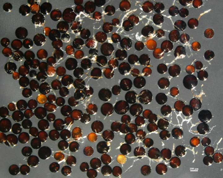
Whole Spores | ||
|---|---|---|
|
|
|
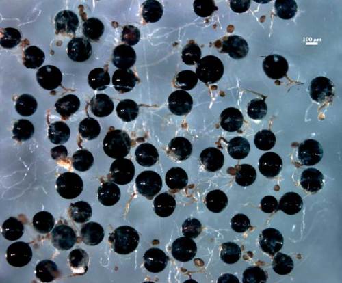
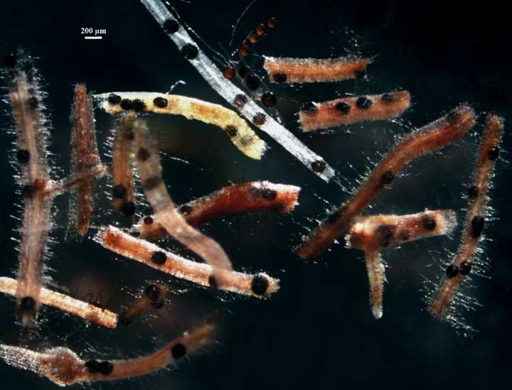
COLOR: Fairly wide range, from red-brown to almost black
SHAPE: Globose to subglobose, rarely irregular.
SIZE DISTRIBUTION: xx µm (mean = ). Spores typically are formed in the rhizosphere, but also can form within roots. This habit varies greatly between strains and even successive cultures of a particular strain
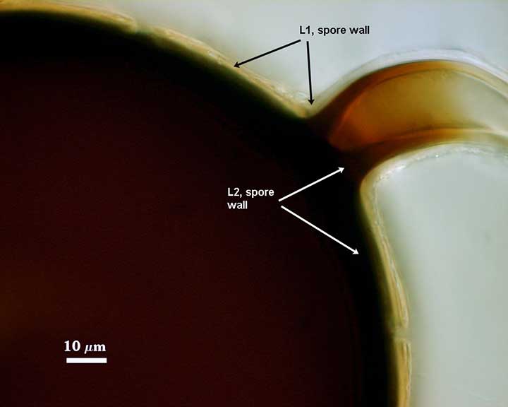
Subcellular Structure of Spores
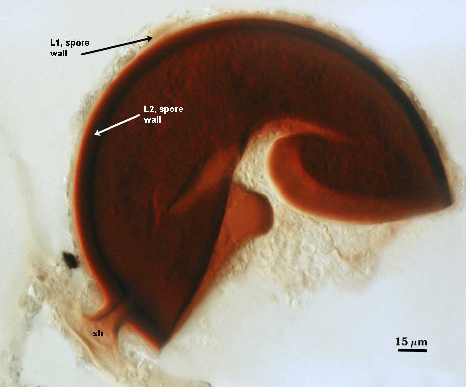
SPORE WALL: Two layers (L1, L2), with the outer layer adherent until it degrades and sloughs.
L1: A hyaline layer; 2-4 µm thick; does not react in Melzer’s reagent. In aged spores, this layer usually is missing except perhaps at the base in the region of the subtending hypha.
L2: An orange-brown to dark red-black laminate layer, 5-9 µm which is continuous with the inner layer of a persistent subtending hypha.
| Smashed Spore | |
|---|---|
|
|
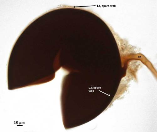
Subtending Hypha
SHAPE: Cylindrical to constricted (see both types above), occasionally slightly flared. Even though this species was named for the constricted phenotype, it only occurs in some percentage of spores (varying with strain).
WIDTH: xx µm at spore.
WALL STRUCTURE: Two layers (L1, L2) that are continuous with the two layers of the spore wall. The outer hyaline layer (L1) often sloughs in older spores, much like it does on the spore surface, so it varies in thickness, rarely exceeding 2-3 µm at the juncture with the spore. The inner layer (L2) is concolorous with the same layer in the spore wall, x-x µm thick at the spore base and then thinning.
OCCLUSION: Varies from a plug to a septum.
The images below can be uploaded into your browser by clicking on the thumbnail or can be downloaded to your computer by clicking on the link below each image. Please do not use these images for other than personal use without expressed permission from INVAM.
High Resolution Images | |
|---|---|
 |  |
 | |