Septoglomus deserticola
(reference accession CA113)
Whole Spores | |
|---|---|
|
|
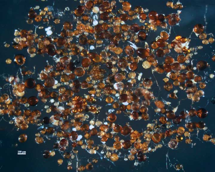
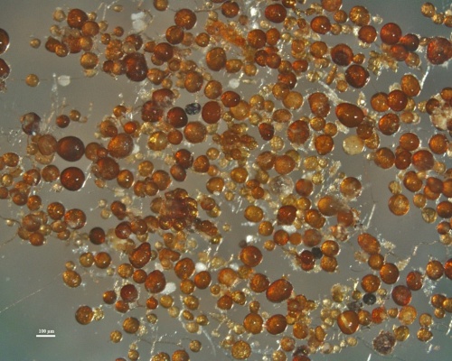
COLOR: orange-brown to dark red-brown. Most are a reddish-brown (20/80/100/0).
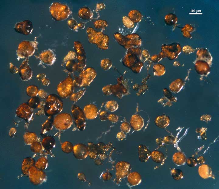
SHAPE: Globose to subglobose usually, but sometimes irregular (see right photo above).
SIZE DISTRIBUTION: Highly variable, from 60-140 µm. Spores typically are formed on very robust hyphae around the root, so they aren’t dislodged as easily as many other glomoid species.
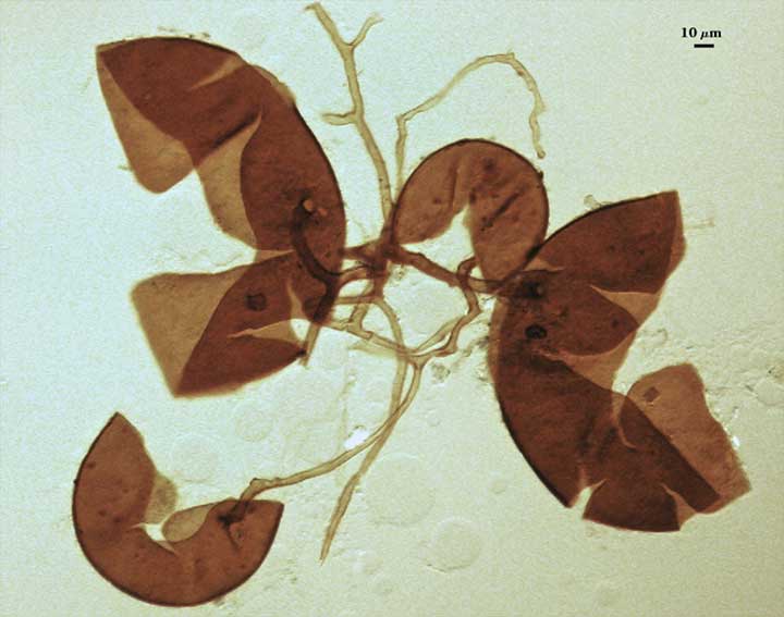
Subcellular Structure of Spores
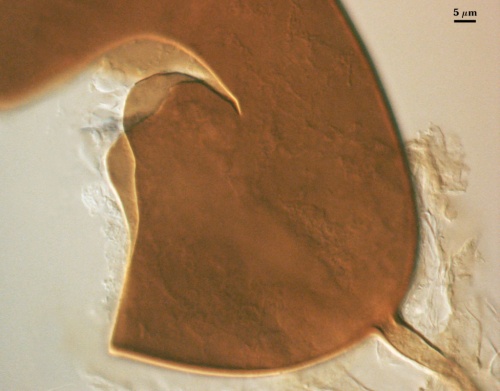
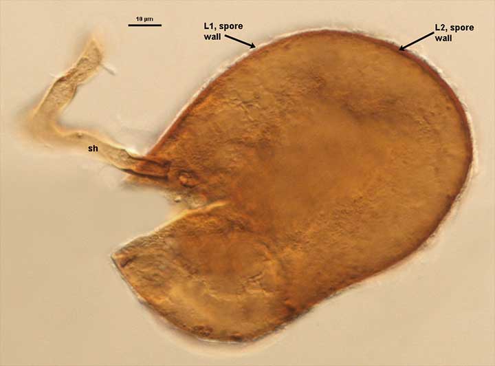
SPORE WALL: Two layers (L1, L2), with the outer layer adherent until it degrades and sloughs.
L1: A hyaline layer; 2-4 µm thick; does not react in Melzer’s reagent. In aged spores, this layer is almost always missing.
L2: An yellow-brown to orange-brown laminate layer, 4-6 µm thick, which is continuous with the inner layer of a persistent subtending hypha. In Melzer’s reagent, this layer produces a dextrinoid reaction (appearing darker red-brown)
| In PVLG | In Melzer's Reagent |
|---|---|
|
|
Subtending Hypha
COLOR: Cylindrical to slightly flared.
WIDTH: 8-12 µm at base of spore.
WALL STRUCTURE: Two layers (L1, L2) that are continuous with the two layers of the spore wall. The outer hyaline layer (L1) is very thin (1 µm or less). The inner layer (L2) is concolorous with the same layer in the spore wall, 1-3 µm thick at the spore base and then thinning to < 2 µm. The hyphae do not break easily, so that spores often are clustered together very loosely (see photo) or are closely associated with root fragments.
| Deserticola | |
|---|---|
|
|
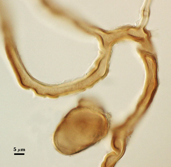
OCCLUSION: Usually a septum.
The images below can be uploaded into your browser by clicking on the thumbnail or can be downloaded to your computer by clicking on the link below each image. Please do not use these images for other than personal use without expressed permission from INVAM.
High Resolution Images | |
|---|---|
 |  |
 |  |