Dentiscutata erythropa
(reference accession MA453B)
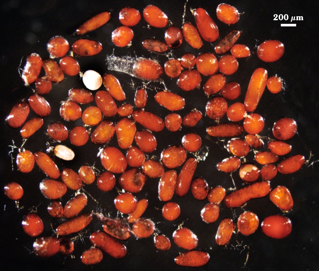
Whole Spores
| Rice Grain | |
|---|---|
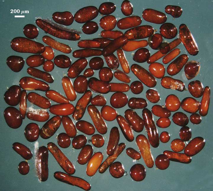 | 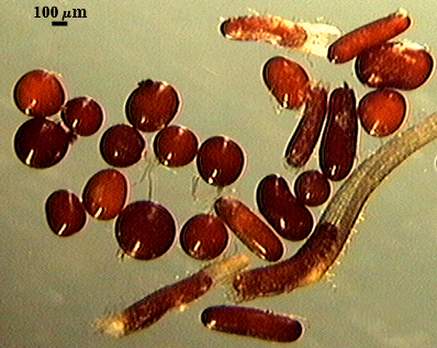 |
COLOR: Red-brown (20-80-60-0) to dark red-brown (20-80-100-10).
SHAPE: Subglobose to oblong, with the latter more common (many spores form within roots).
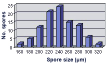
SIZE DISTRIBUTION: 160-320 µm, mean = 240 µm (n = 100).
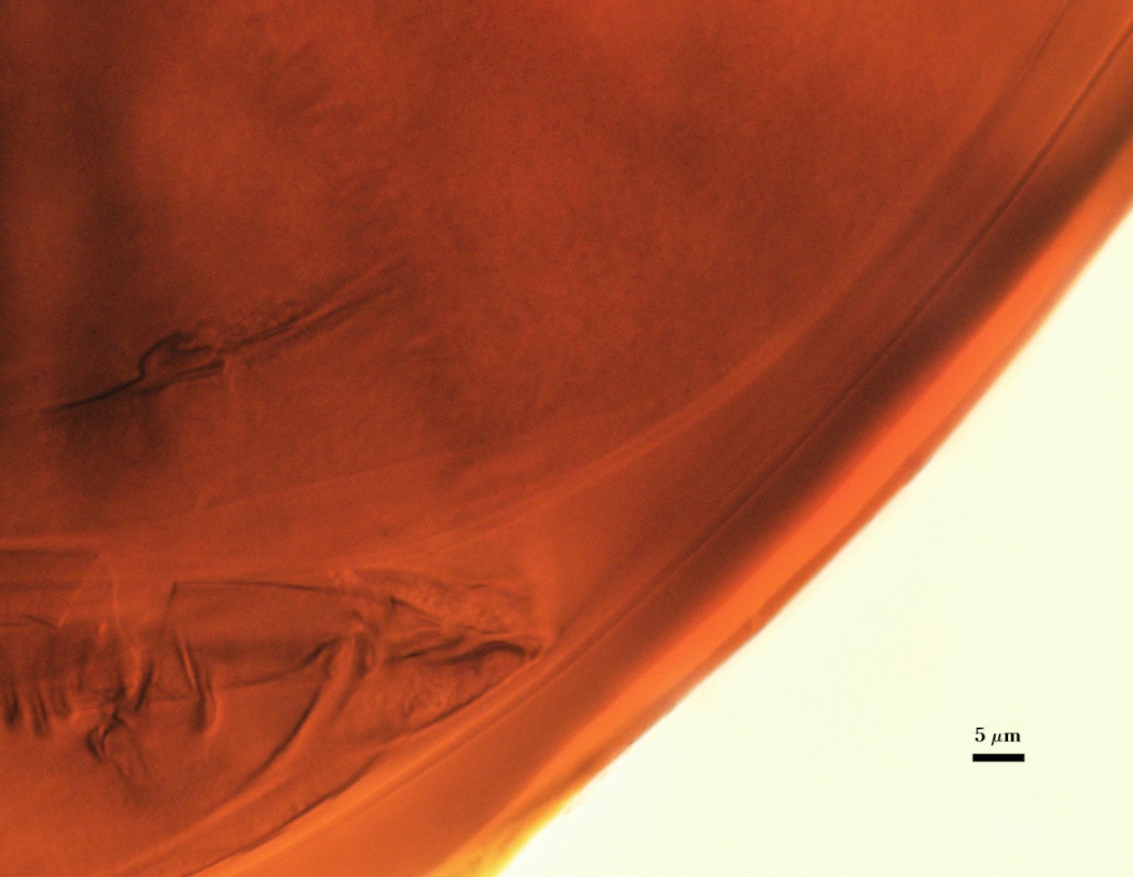
Subcellular Structure of Spores
SPORE WALL: Two layers (L1 and L2) that are adherent that in juvenile spores are of equal thickness, with the laminate layer thickening as the spore wall is differentiated.
L1: An outer permanent rigid layer with a smooth surface, dark red-brown (20-60-100-10), 0.8-1.2 µm thick. Tightly adherent to L2.
L2: A layer consisting of dark orange (20-80-80-0) to red-brown (20-80-100-0) sublayers (or laminae) that increase in number with thickness; 3.2-7.4 m (mean of 6.8 m) thick in mature spores.
| In PVLG | ||
|---|---|---|
 |  | |
| In PVLG and Melzer's reagent (1:1 v/v) | ||
|---|---|---|
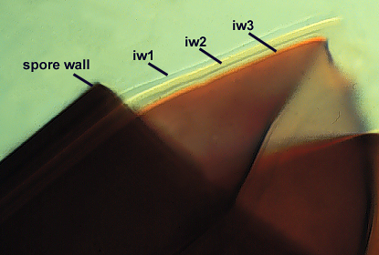 |  | 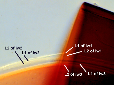 |
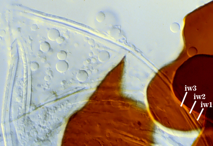
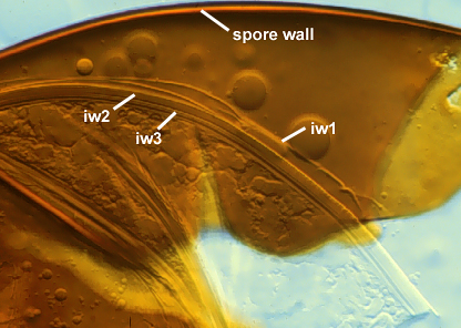
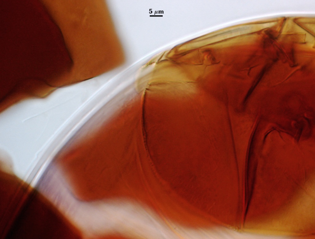
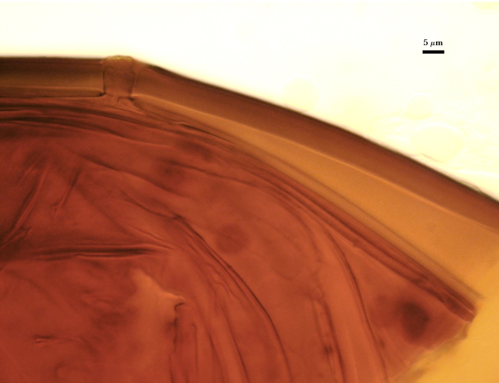
GERMINAL WALLS: Three flexible hyaline inner walls (gw1, gw2, and gw3), all of which tend to be adherent to each other (see photos above and closer details in the sequence below).
GW1: Two adherent layers (L1 and L2) are formed. L1 is less than 0.5 µm thick; L2 is slightly thicker (0.5-0.8 µm). Both layers are so thin that when they are adherent, they appear as one layer. This layer often is hard to detect because it often is tightly adherent to IW2 or breaks up when a spore is broken.
GW2: Two adherent layers (L1 and L2) are formed. L1 is less than 0.5 µm thick; L2 is 1.2-3.2 µm thick and becomes slightly discolored (0-0-20-0) in Melzer’s reagent.
GW3: Two adherent layers (L1 and L2) are formed. L1 is 0.8-2.4 µm thick; L2 is 0.9-1.6 µm thick and stains pinkish (0-40-20-0) to pinkish purple (0-60-30-10).
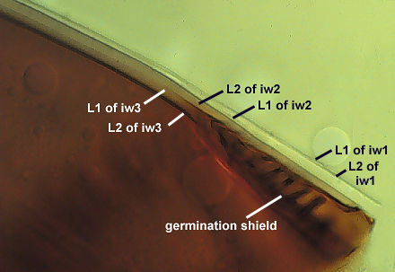
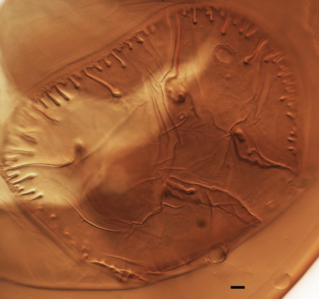
| Germination wall lines |
|---|
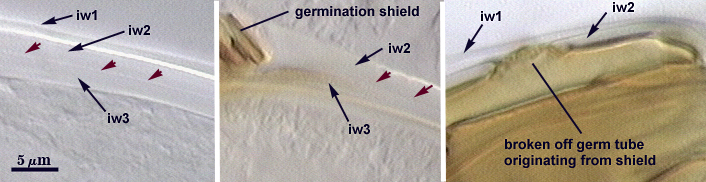 |
| Left to right: (i) detail of the three germinal walls, (ii) position of shield between gw2 and gw3, (iii) remnant scar of germ tube pushing through gw1 and gw2 (and the spore wall) |
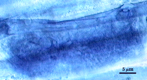
Subtending Hypha
WIDTH OF SPOROGENOUS CELL: 38-50 µm (mean = 45 µm).
SPOROGENOUS CELL WALL: Two layers (L1 and L2) probably are present (continuous with the two layers of the spore wall), but only L2 is readily discernible at the level of the compound microscope.
L2: Yellow-brown in color (0-10-60-0), 2.4-4.8 µm thick near the spore and then thinning to 1.0-1.5 µm beyond the sporogenous cell.
OCCLUSION: Closure by a plug concolorous with L2 of the spore wall.
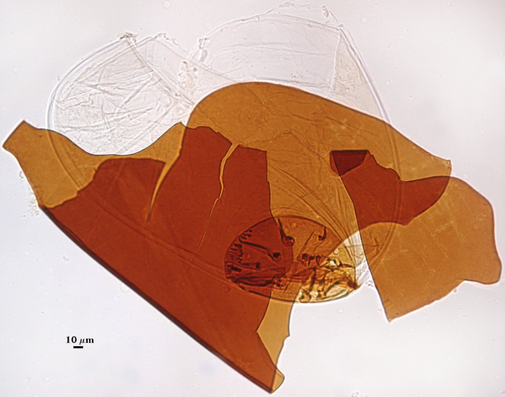
Germination
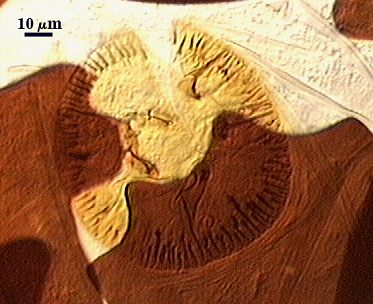
COLOR: Yellow brown (0-10-100-10)
SHAPE: Oblong, with length approximately 1.5 times that of the width. Position of the shield is on the innermost germinal wall (gw3). Margins of the shield are deeply invaginated (2-30 µm deep), with some bifurcating at the tips. The outer boundary of the shield is very robust (shields rarely break apart in broken spores), 2.5-3 µm thick.
SIZE: Generally 45-90×95-210 µm.
Auxiliary Cells
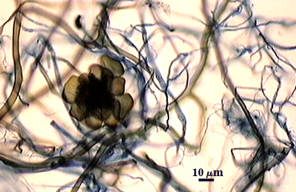
Aggregate (2-15) cells borne on coiled brown (20-40-80-0) hyphae 5-7 µm in diameter; thin-walled (< 1 µm thick), with wall pale yellow (0-0-5-0) in transmitted light; each cell with tuberculate surface, with swellings 1.8-3.2 µm wide and 8.6-12 µm high.
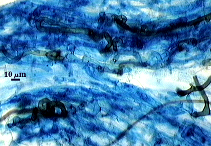
Mycorrhizae
Intraradical arbuscules and hyphae consistently stain darkly in roots treated with trypan blue. Arbuscules with thick trunk and often short coils (6-8 µm in diameter) and many finely tipped branches. Hyphae often with knobs or projections, 5-10 µm in diameter, usually densely coiled near and around entry points but also widely distributed throughout the root cortex. Extraradical hyphae consisting of very fine hyphae (< 2 µm in diameter) and coarser dark brown (20-40-100-0) hyphae 3-8 µm in diameter (see hyphae associated with auxiliary cell cluster in photo above).
| Arbuscules in cortical cells of corn roots | ||
|---|---|---|
 | 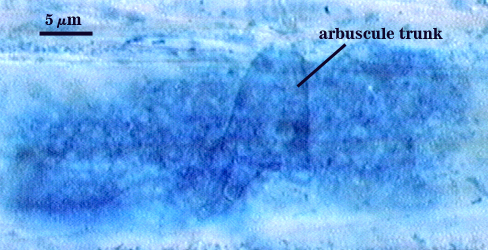 | 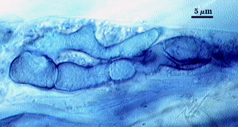 |
| Other structures of mycorrhizae in corn | ||
|---|---|---|
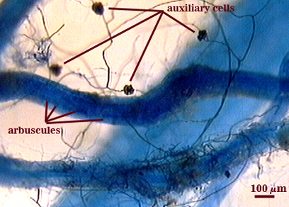 | 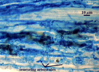 |  |
Notes
Some spores are formed within the root cortex, and may account in part for the large proportion of oblong spores. Since many soil-borne spores are of similar shape, this trait also appears to be heritable (under genetic control). Spores in dried pot culture inoculum are not as long-lasting as their lighter colored relatives, rarely extending longer than 12 months. In our experience, many spores collected from field soils contain internal parasites, even though they appear healthy when examined intact under a dissecting microscope.
This species was described before Scutellospora was separated from Gigaspora. Koske and Walker (1984) described spores as having an inner thin “unit wall” (probably gw1) and an inner “laminate wall” (which actually consists of gw2 and gw3 when tightly adherent as exemplified in the photo sequence above). The difficulties in interpretation of germinal wall structure and organization was understandable at the time this species was first described, since little was known of kinds and properties of these walls or of the immutable position of the germination shield on the innermost germinal wall (regardless of how many of these inner walls are present). The two innermost germinal walls are easiest to distinguish, despite their tight adherence, because they separate where a germination shield forms (see photos above).
The images below can be uploaded into your browser by clicking on the thumbnail or can be downloaded to your computer by clicking on the link below each image. Please do not use these images for other than personal use without expressed permission from INVAM.
High Resolution Images | |
|---|---|
 |  |
 |  |
 |  |
Links to Gene Sequences in Genbank
Reference
Koske, R. E. and C. Walker. 1984. Gigaspora erythropa, a new species forming arbuscular mycorrhizae. Mycologia 76:250-255.