Claroideoglomus etunicatum
(reference accession NE108A)
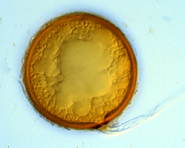
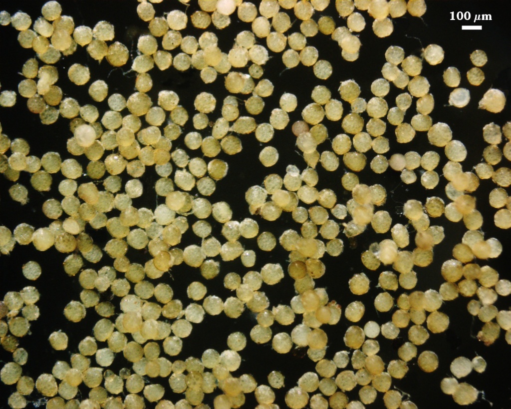
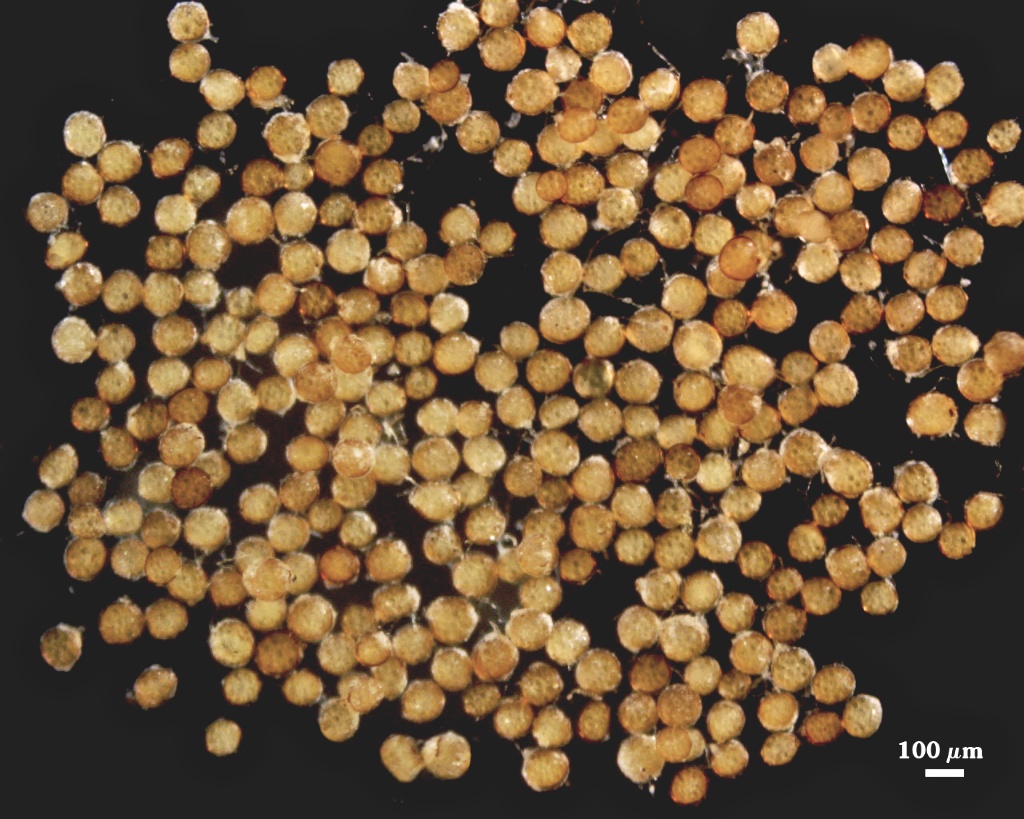
Whole Spores
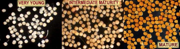
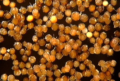
COLOR: Orange (0-10-90-5) to red brown(0-60-100-0).
SHAPE: Globose, subglobose.
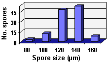
SIZE DISTRIBUTION: 60-160 µm, mean = 129 µm (n = 120).
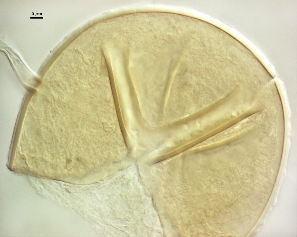
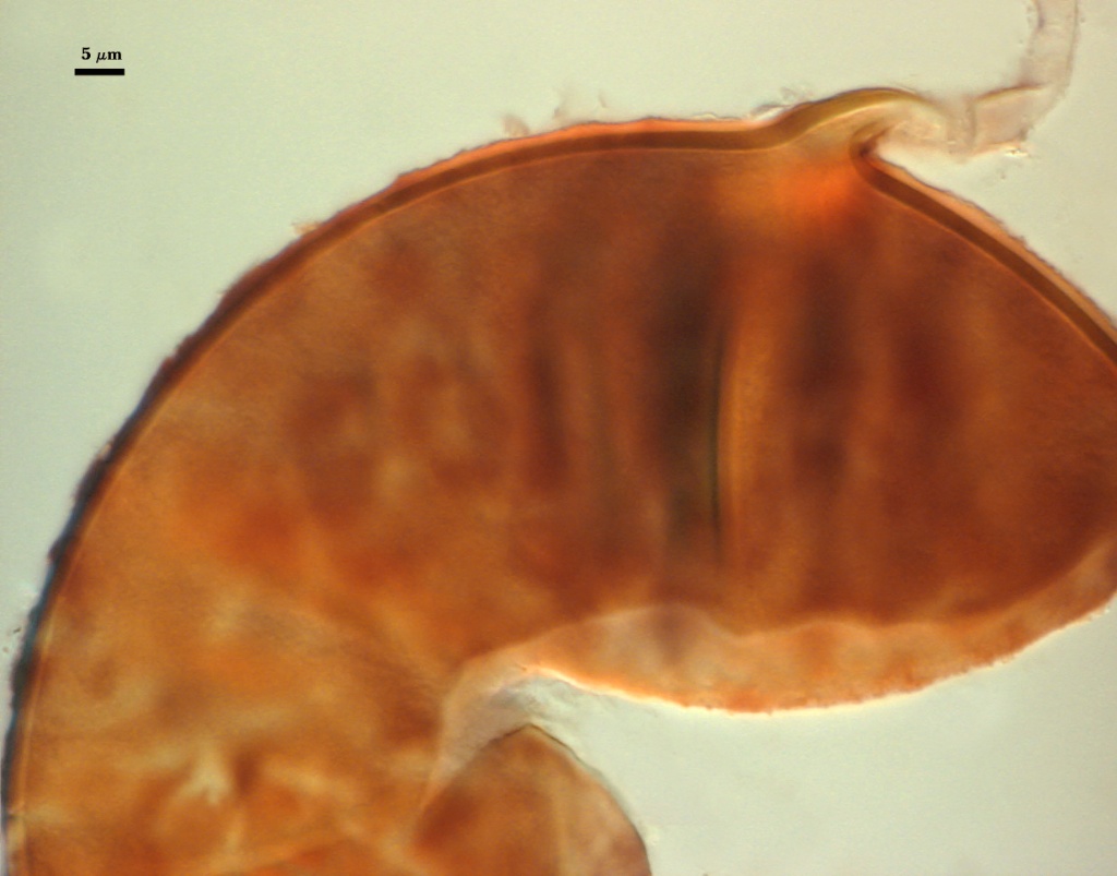
Subcellular Structure of Spores
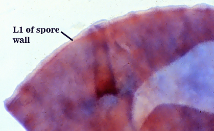
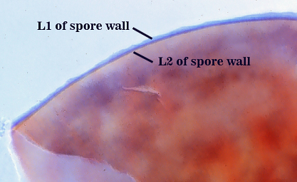
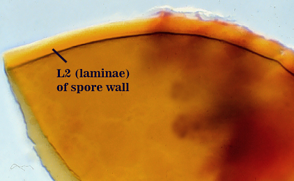
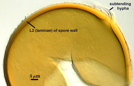
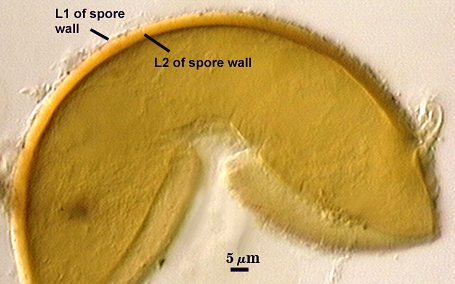
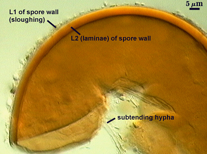
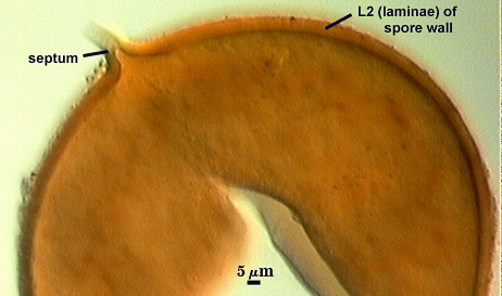
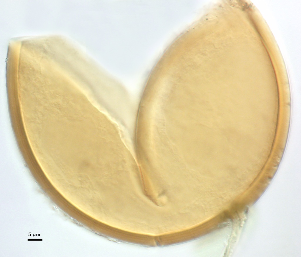
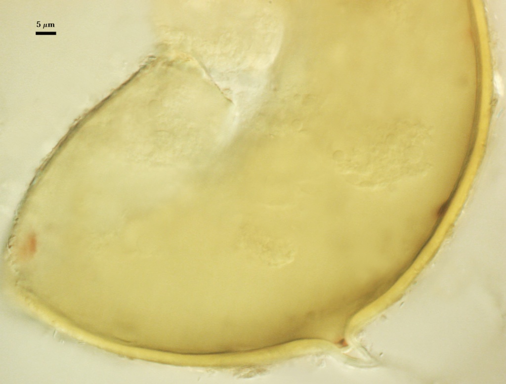
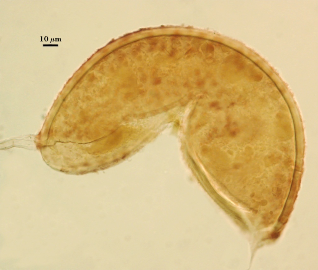
SPORE WALL: Consisting of two layers (L1 and L2) that differentiate consecutively as spores develop. Initially, only L1 is present in both the spore and subtending hyphal wall. L2 begins as a single layer and then thickens with formation of additional sublayers of the same phenotypic properties.
| Sequential stages in differentiation of the spore wall, from left to right. All spores mounted in Melzer’s reagent | |
|---|---|
 |  |
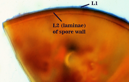 |  |
L1: An outer layer; mucilagenous (having some plasticity with uneven outer surface); staining pink (0-60-30-10) to reddish purple (20-80-60-0) in Melzer’s reagent; 0.6-2.8 µm thick in young spores. This layer degrades and sloughs as spores age, so that it may be present in patches (usually detected in Melzer’s reagent) or appear as a granular layer. This layer appears to be a substrate for bacteria, and spores will acquire a distinct halo when stored in tap water several days at room temperature and several weeks in a refrigerator.
L2: A layer consisting of thin adherent sublayers (or laminae), light orange-brown (0-20-60-0) to red-brown (0-40-100-10) in color; 4.4-6.4 µm thick (mean of 5.3 µm). Sublayers in the region of the subtending hypha sometimes thicken to 7.6 µm.
| In PVLG | ||
|---|---|---|
|
|
|
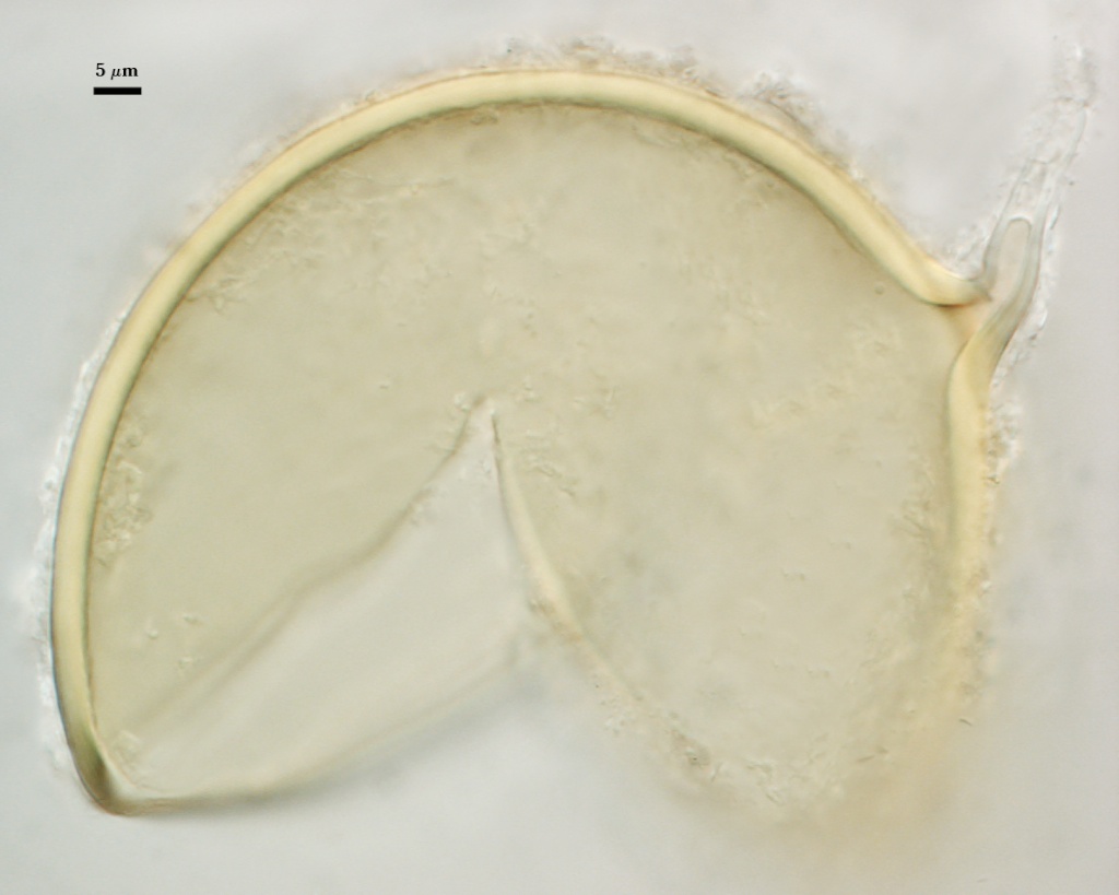
| Spores mounted in PVLG and Melzer’s reagent (1:1 v/v) | |
|---|---|
|
|
Subtending Hypha
SHAPE: Cylindrical to slightly flared (see photos above).
WIDTH: 5-10.2 µm (mean = 7.9 µm).
COMPOSITE WALL THICKNESS: < 1-2 µm.
WALL STRUCTURE: Two layers (L1 and L2) continuous with the two layers of the spore wall.
L1: The outer layer, usually present only near point of attachment of mature spores; always present on juvenile hyphae attached to juvenile spores. L2: Although is layer is continuous with L2 of the spore wall, it is pale yellow (0-0-10-0) to pale orange (0-0-20-0) from the region of attachment to the spore.
OCCLUSION: Innermost sublayer of the laminate layer of the spore wall; in some spores, this bridging structure resembles a septum (see photos above).
Germination
A germ tube emerges from the lumen of the subtending hypha.
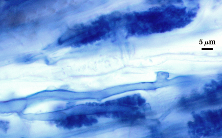
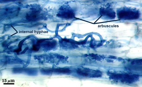
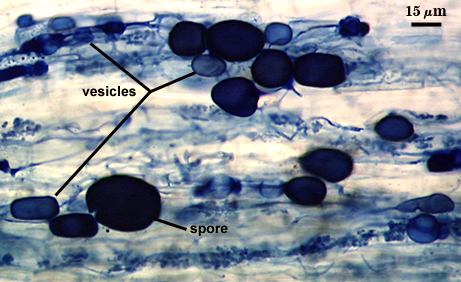
Mycorrhizae
Numerous arbuscules and vesicles produced within the first eight weeks of culture on sudangrass. Hyphae form long infection units with hyphae interconnected by perpendicular branches (“H” or “h” connections). Stain darkly in trypan blue.
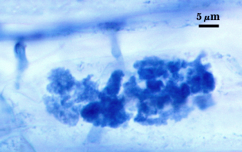
| Arbuscules in corn roots | |
|---|---|
|
|
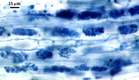
| All mycorrhizal structures in corn roots | ||
|---|---|---|
|
|
|
Notes
Young spores are white, changing to a tan color (0-20-60-0) as the laminate layer (L2) of the spore wall thickens and finally to the mature state (see above). This species is one of the most common found throughout the world, based on the number and distribution of accessions in INVAM (from tundra of Alaska to deserts of Namibia).
The images below can be uploaded into your browser by clicking on the thumbnail or can be downloaded to your computer by clicking on the link below each image. Please do not use these images for other than personal use without expressed permission from INVAM.
High Resolution Images | |
|---|---|
 |  |
 |  |
 |  |
 |  |