Ambispora gerdemannii (acaulosporoid synanamorph)
Whole Spores
| Acaulosporoid Synanamorph | |
|---|---|
|
|
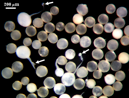
 COLOR: Cream (0-0-15-0) to dark orange-brown (0-40-100-10), most orange-brown (0-30-80-0)
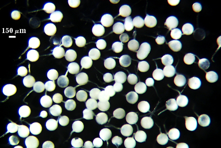
SHAPE: Globose, subglobose.
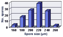
SIZE DISTRIBUTION: 160-260 µm, mean = 213 µm (n = 116)
Subcellular Structure of Spores
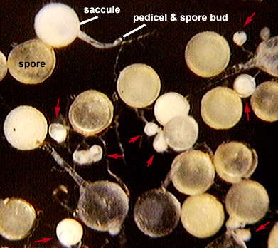
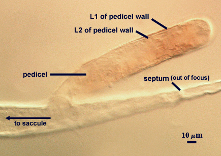
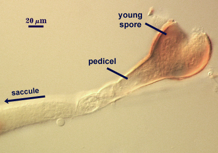
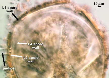
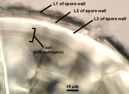
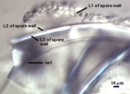
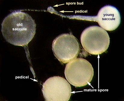
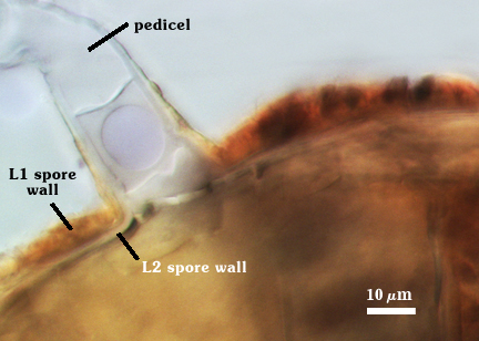
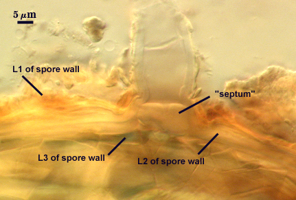
SPORE WALL: Consisting of three layers (L1, L2 and L3) in which L2 branches from the hypha subtending the “sporiferous saccule” (termed a pedicel), followed by synthesis of the outermost layer, L1 (time of synthesis easily pinpointed by sudden appearance of red coloration caused by reaction of L1 in Melzer’s reagent—see second and third photos from left below). L1 originates on the pedicel and forms continuously on the spore; it thickens only on the spore as it differentiates further. In contrast, L2 thickens on both pedicel and spore as ontogenesis progresses. L3 arises inside L2 and appears to grow from it (some evidence of association by emergence from the pedicel).
| Sequence (left to right – click to enlarge) in the differentiation of a spore from a pedicel branching from the neck of a “sporiferous saccule”. | ||
|---|---|---|
|
|
|
|
|
|
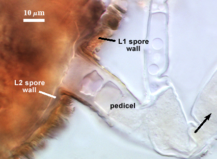
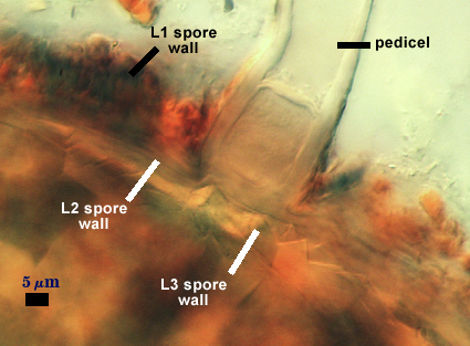
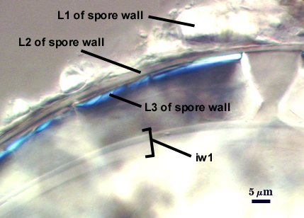
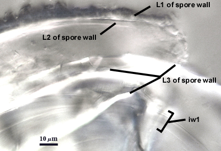
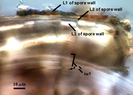
L1: A friable layer with deep fissures in the surface that breaks apart easily with applied pressure, expanding somewhat in PVLG-based mountants; hyaline in youth, becoming pale orange-brown (0-20-40-0) with age; 6-15 µm thick when intact on the spore wall; degrading and/or sloughing with age; staining dark orange-brown (0-60-100-10) to red-brown (20-60-100-0) in Melzer’s reagent.
L2: Hyaline, continuous with the wall of the pedicel; 2.5-4 µm thick; consisting of sublayers that usually are adherent, but which sometimes separate slightly. The entire layer is somewhat pliable and may slough (with subtending pedicel) together with L1.
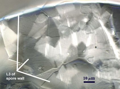
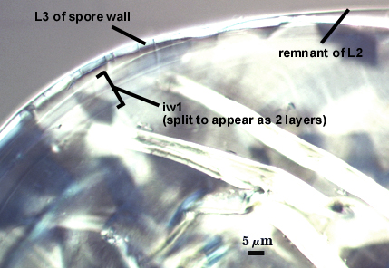
L3: Hyaline, rigid, 1.8-2.5 µm thick (most 2 µm), having slate-like properties when broken, forming numerous fracture lines and polygonal shards rather than a clean break (see photo below). This layer has birefringent properties in polarized light so that light is reflected brightly, especially at fracture edges. It is the extensive breakup and concomitant light-scattering of this layer in field collected spores which made this species so hard to describe (see notes section below).
iw: Hyaline, semi-pliable wall, 3-7 µm thick (mean = 5.2 µm); consisting of multiple sublayers (see photos below) that can split into three components: a thin outer and inner layer and a thick middle layer; no reaction in Melzer’s reagent.
| In PVLG | |
|---|---|
|
|
|
|
| In Melzer’s reagent | |
|---|---|
|
|
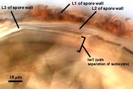
Spores with outer two layers sloughed (photos below) are bright white and spores with all layers present (photos above) are pale to dark brown.
|
|
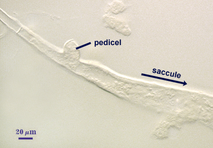
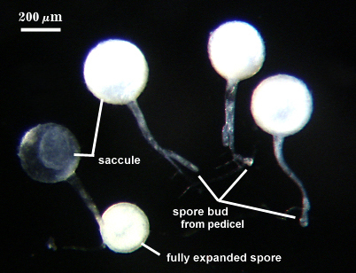
Pedicel
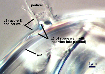
PEDICEL: So named because it is a feature divergent from most Acaulospora species, arising as a branch from the neck of the “sporiferous saccule”, 200-280 µm from the “saccule”. The pedicel is 65-90 µm in length (saccule hypha to spore), 16-28 µm wide at the base of the spore. Just prior to formation a distal thin septum forms that closes off contents of the pedicel and “saccule” from parent hyphae (see photos in developmental sequence above). The septum is 1-2 µm thick and forms 38-55 µm from the saccule.
PEDICEL WALL: Two hyaline layers (L1 and L2). The outer layer (L1) is continous with L1 of the spore wall, 1.5-2 µm thick, staining dark orange-brown (0-60-80-0) in Melzer’s reagent, sloughing except near the spore base as the spore differentiates. The inner layer (L2) is permanent and continuous with L2 of the spore wall, 3-6 µm thick at the base of the spore when it is mature.
Occlusion
The middle layer of the spore wall (L2) bridges the pedicel lumen, distinguished as a flat hyaline “septum” 4-10 µm thick.
| Middle Layer | |
|---|---|
|
|
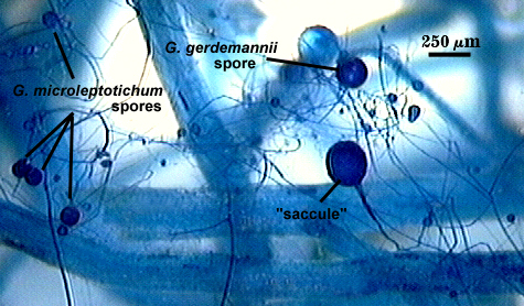
Sporiferous Saccule
| Acaulosporoid Synanamorph | ||
|---|---|---|
|
|
|
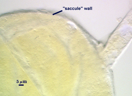
COLOR: White in youth (see far left photo above) to hyaline at maturity.
SHAPE: Globose to subglobose (see far right photo above for unusual behavior in producing protrusions)
SIZE DISTRIBUTION: 220-300 µm, mean = 260 µm (n = 96)
WALL STRUCTURE: Hyaline and of variable thickness due to patchy sloughing of material so that the outer surface appears flaky; 3.5-7 µm thick.
SUBTENDING HYPHA: 28-56 µm wide at the saccule, with a hyaline wall 2.5-4 µm thick.
Germination
Not yet observed in in vitro assays.
Mycorrhizae
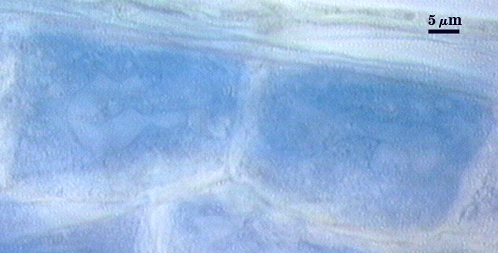
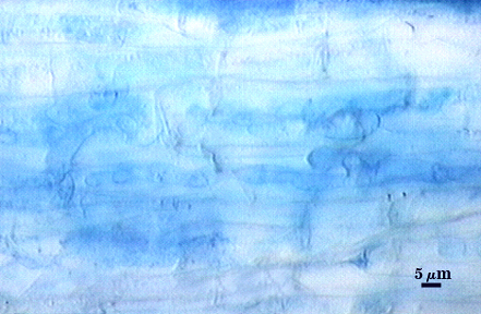
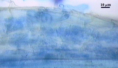
Coiled and knobby hyphae, 4-9.0 µm in diameter, with finely branched arbuscules in cortical cells. No vesicles have been observed in INVAM accessions examined to date. The staining reaction of fungal structures in Melzer’s reagent is highly variable, from invisible to a very faint pale blue that often is hard to detect under a stereomicroscope (mounted roots are better). Contrast was digitally enhanced to view structural details that often are hard to see clearly otherwise.
| Arbuscules in corn root cortical cells | ||
|---|---|---|
 |  |  |
Notes
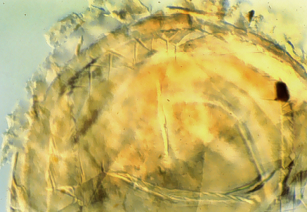
The description and classification of Glomus gerdemannii by Rose et al. (1979) was based on appearance of field-collected spores. The unusual properties of L3 of the spore wall when broken caused the spores to look so “messy” that interpretation of structure was difficult (see photo of holotype specimen below). They did not appreciate at that time the extent to which spore morphology could be altered by senescence, parasitism, and unnumerable other variables in soil, especially in species producing spores with such a complex spore and inner wall organization.
Mode of saccule and spore formation, as well as spore size and subcellular organization, are identical to that of the acaulosporoid synanamorph of Am. leptoticha. In fact, spores of the two species, with or without attachment to the “sporiferous saccule”, are almost indistinguishable under a stereomicroscope. The sole difference in spore morphology resides in properties of the inner two layers of the spore wall (L2, L3). These layers are smooth, with the inner layer breaking into shards with pressure in spores of Am. gerdemannii, whereas these two layers have hemispherical depressions/protrusions that fit together like two pieces in a jigsaw puzzle in spores of Am. leptoticha.
Reference
- Rose, S., B. A. Daniels, and J. M. Trappe. 1979. Glomus gerdemannii sp. nov. Mycotaxon 8:297-301.