Ambispora gerdemannii (glomoid synanamorph)
Whole Spores
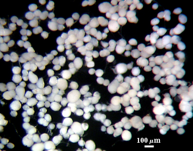
 COLOR: Hyaline to white to very pale yellow (0-0-5-0)
SHAPE: Globose, subglobose, often irregular.
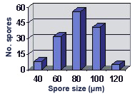
SIZE DISTRIBUTION: 40-120 µm, mean = 80 µm (n = 142)
Subcellular Structure of Spores
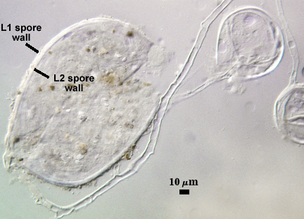
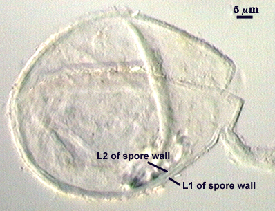
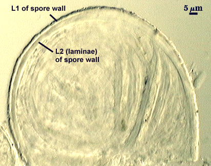
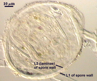
SPORE WALL: Two layers (L1 and L2) which usually are adherent.
| In PVLG | |
|---|---|
|
|
| In Melzer’s reagent | |
|---|---|
|
|
L1: Outer layer, hyaline to very pale yellow (0-0-10-0), with a flaky surface, < 0.5-1 µm thick, not reacting visibly with Melzer’s reagent when intact. This layer tends to break down with age and sloughs to varying degrees. Sometimes, this layer accumulates some organic debris, giving the spore surface a “dirty” appearance.
L2: A layer consisting of hyaline to subhyaline (0-0-10-0) sublayers (or laminae) that increase in number with thickness. The composite layer is 1.5-4 µm thick, and appears to have some resiliency when pressure is applied. Thus, as spores are broken, this layer may “flatten” or fold instead of breaking (especially in younger spores), and measure a thickness up to 6 µm. The inner sublayers of L2 may appear wrinkled within minute folds because of this resiliency, suggesting (erroneously) the presence of a very thin flexible layer (or “membranous wall”).
Subtending Hypha
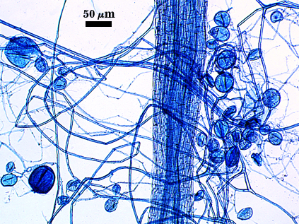
SHAPE: Cylindrical to slightly flared (see photos above).
WIDTH: 6-12 µm at the juncture of the spore wall
COMPOSITE WALL THICKNESS: 1-2 µm
HYPHAL WALL: Two layers (L1 and L2) continuous with the two layers of the spore wall (see photos above). Both layers are of near equal thickness, 0.5-1 µm. Thickness of the hypha wall thins to < 0.5 µm some distance from the spore.
Occlusion
Thickening of the inner layer (L2) of the spore wall.
Germination
Not yet observed in in vitro assays.
Notes
Spores are identical in mode of formation and subcellular structure of the glomoid synanamorph of Ambispora leptoticha. The only morphological difference is the size range of spores. Spores of this morphotype do not exceed 120 µm, whereas those of the Am. leptoticha morphotype can grow to 260 µm.
