Ambispora leptoticha (glomoid morph)
Whole Spores
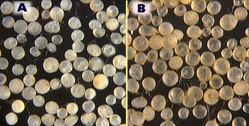
 COLOR: White to pale yellow brown (0-5-30-0)
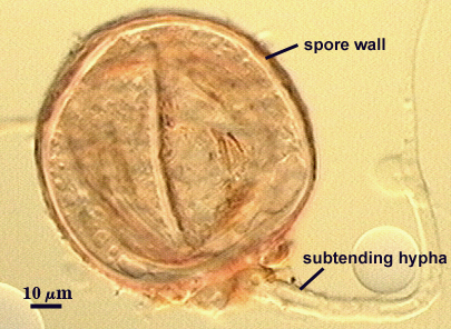
SHAPE: Globose, subglobose, with many spores irregular.
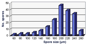
SIZE DISTRIBUTION: 60-260 µm
Subcellular Structure of Spores
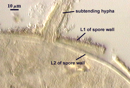
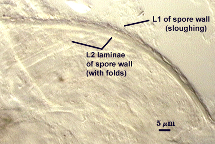
SPORE WALL: Two layers (L1 and L2), often adherent but sometimes separating; both colorless in youth.
| Spores mounted in PVLG | |
|---|---|
|
|
| Spores mounted in Melzer’s reagent | ||
|---|---|---|
|
|
|
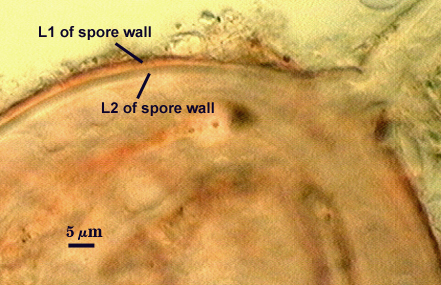
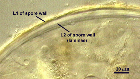
L1: Outer layer, hyaline to pale yellow (0-0-20-0), with a flaky surface to which bits of debris often are attached. It is this characteristic which often gives whole spores a “dirty” appearance and additional brown coloration. This layer tends to break down with age and thus often is of variable thickness, ranging usually from 2.4-4 µm. The surface of this layer also reacts in Melzer’s reagent, with the color ranging from a pale pink (0-15-20-0) to orange-red (0-60-60-0).
L2: A layer consisting of hyaline to subhyaline (0-0-10-0) sublayers (or laminae) that increase in number with thickness. The composite layer is 4-8 µm thick, and appears to have some resiliency when pressure is applied. Thus, as spores are broken, this layer may “flatten” or fold instead of breaking (especially in younger spores). The inner sublayers of L2 often appear wrinkled within minute folds because of this resiliency. Despite this appearance, there are no flexible inner walls present in mature spores.
Subtending Hypha
SHAPE: Cylindrical, but more often flared (see photos above).
WIDTH: 15-22 µm (mean = 18.6 µm)
TOTAL WALL THICKNESS: 4-6 µm
WALL STRUCTURE: 2 layers (L1 and L2) continuous with the two layers of the spore wall (see photos above).
L1: The outer layer, rarely extending more than 20 µm down the length of the subtending hypha in fully mature spores, 1.5-3 µm thick near spore.
L2: The layer which gives the mature subtending hypha structural integrity. It is thickest near the spore (2-3.2 µm) and then thinning to 1.5 µm or less.
OCCLUSION: Closure by thickening of the spore wall in the region of hyphal attachment.
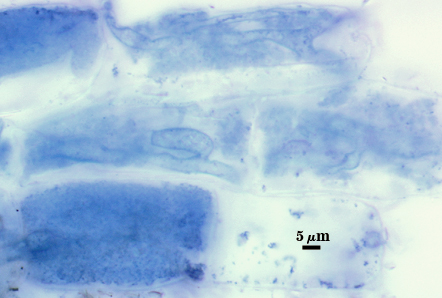
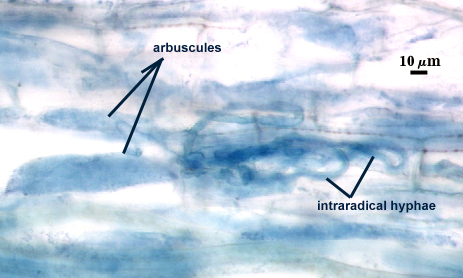
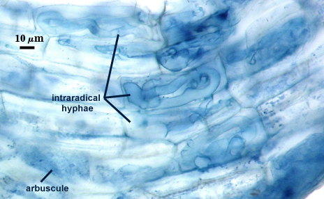
Mycorrhizae
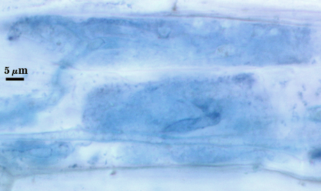
Arbuscules and hyphae in cortical cells of sorghum, sudangrass, corn and red clover roots stain with variable intensity in trypan blue, ranging from almost invisible, a pale blue (most common), to slightly dark blue. Hyphae often are coiled within individual cells, with similar dimensions (8-12 µm wide) inside and outside roots. Contrast in photos is digitally enhanced to better see mycorrhizal structures.
| Mycorrhizal structures | |||
|---|---|---|---|
|
|
|
|
Notes
Spores range so much in size because those in the lower range (35-60 µm) are produced abundantly on the same hypha as the larger spores. These smaller spores resemble “vesicles” except that they have a similar spore wall structure (only the layers are thinner).