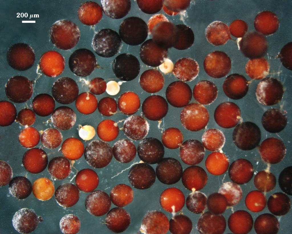
Racocetra gregaria
(reference accession BR239)
Whole Spores
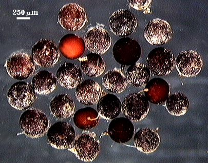
 COLOR: Red-brown (20-80-100-10) to dark red-brown (40-80-80-0)
SHAPE: Globose to subglobose
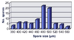
SIZE DISTRIBUTION: 380-520 µm, mean = 473 µm (n = 82)
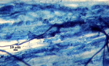
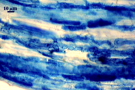
Subcellular Structure of Spores
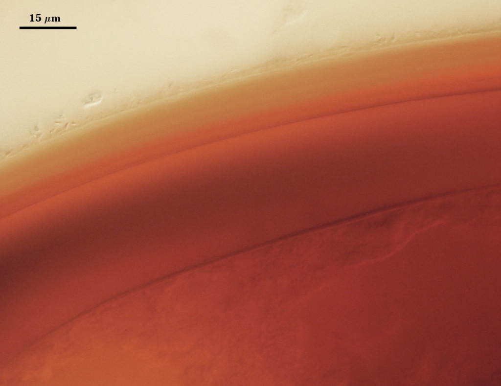
SPORE WALL: Two layers (L1 and L2) that are adherent that in juvenile spores are of equal thickness, with the laminate layer thickening as the spore wall is differentiated.
| Spores mounted in PVLG | |||
|---|---|---|---|
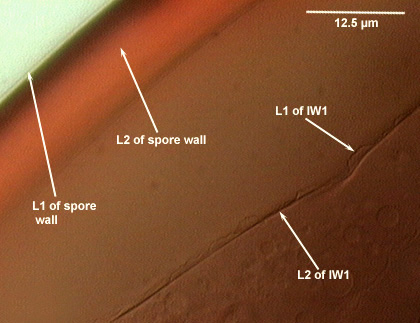 | 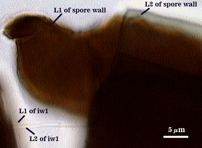 | 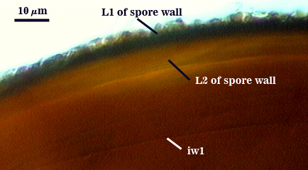 | 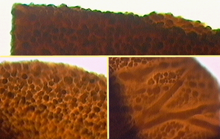 |
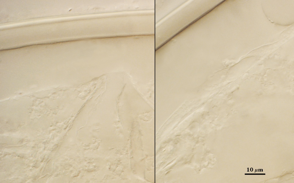
L1: An outer permanent rigid layer, dark brown (60-80-30-10), 0.7-1.8 m thick. The surface of this layer forms ornamentations consisting of rounded warts, most 11-26.7 µm wide x 3-7 µm high.
L2: A layer consisting of sublayers (or laminae) that appear dark brown because of coverage by L1, but appears to be hyaline or pale yellow (see second photo above); 8-12.6 m (mean of 10 µm). This layer stains dark red brown (20-60-40-0) to brown (40-80-80-0) in Melzer’s reagent.
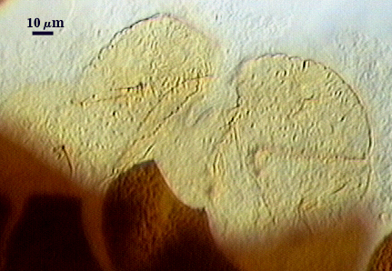
GERMINAL WALLS: One flexible hyaline inner wall (gw1) formed independent of the spore wall and subtending hypha, with two adherent layers.
GW1: Two hyaline layers (L1 and L2) that in field-collected spores are so tightly adherent they appear as one layer (the condition of type specimens used to describe the species). In spores from pot cultures, the outer layer often separates in small folds from parts of the wall giving the wall a blistered appearance. L1: A thin layer less than 0.5 µm thick; thus difficult to resolve without differential interference optics; no reaction in Melzer’s reagent. L2: A slightly thicker layer, 0.6-1.3 µm thick; no reaction in Melzer’s reagent.
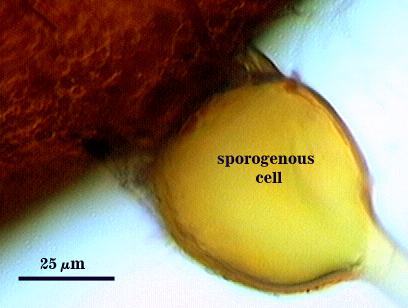
Subtending Hypha
WIDTH OF SPOROGENOUS CELL: 50-66 µm (mean = 57.5 µm)
SPOROGENOUS CELL WALL: Two hyaline layers (L1 and L2) probably are present (continuous with the two layers of the spore wall), but only L2 is readily discernible at the level of the compound microscope.
L2: Yellow-brown (0-20-80-0), 4.0-6.3 µm thick near the spore and then thinning to 1.4-1.6 µm beyond the sporogenous cell.
OCCLUSION: Closure by a plug concolorous with the laminate layer of the spore wall.
Germination
COLOR: Tan (0-10-20-10)
SHAPE: Ovoid, with length approximately 1.5 times that of the width. Margins of the shield with many convolutions that appear as deep warts when viewed from the side. It also has a rugose appearance in plan view that contribute to the warty surface in longitudinal view. Position of the shield is between the spore and germinal wall.
Auxiliary Cells
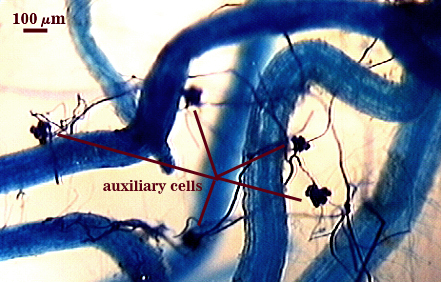
Cells in aggregates of 5-13 (mean = 8), subglobose, ovoid to clavate, borne on coiled hyaline hyphae, thin-walled (< 1 µm thick), light brown (0-20-50-10) in transmitted light, each cell with tuberculate surface, with swellings 1-5 µm high and 3-10 µm wide.
Mycorrhizae
Extraradical hyphae of two morphological types: one wide (3-7 µm) and the other thinner (1.5-2 .0 µm). The former usually is the infective hyphae at entry points and forms knobby swellings there and near auxiliary cells. Intraradical arbuscules and hyphae consistently stain darkly in roots treated with trypan blue. Arbuscular hyphae branch to form many fine tips from a swollen basal hypha. Intraradical hyphae 3-9 µm wide, with knobs, projections and swollen areas (up to 12 µm wide), and usually densely coiled near entry points and in outer cortical cells.
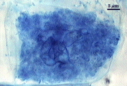
| Arbuscules in corn | ||
|---|---|---|
|
|
|
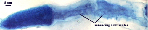
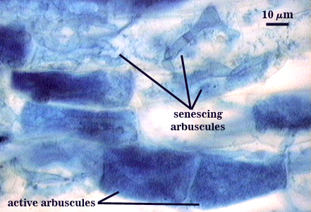
| Other mycorrhizae structures in corn | ||
|---|---|---|
|
|
|
Notes
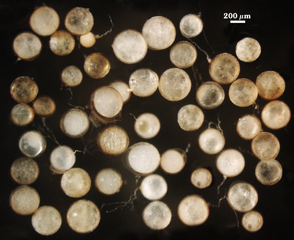
Immature spores are white, then turning bright orange (0-40-100-0) in an intermediate juvenile stage (prior to complete differentiation of the spore wall). Ornamentations on the spore wall are completely formed before any inner wall is synthesized. Both in pot cultures and in the field, many extracted spores are parasitized and do not germinate. In field-collected spores, the spore wall surface (L1) often has tracks or fissures created by microorganisms or microfauna. Increased sensitivity of spores to microbial deterioration prevents cultures from being stored long.
The images below can be uploaded into your browser by clicking on the thumbnail or can be downloaded to your computer by clicking on the link below each image. Please do not use these images for other than personal use without expressed permission from INVAM.
High Resolution Images | |
|---|---|
 |  |
 |  |