Dentiscutata Heterogama Albino Mutant
(reference accession IL203)
Whole Spores
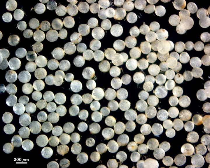
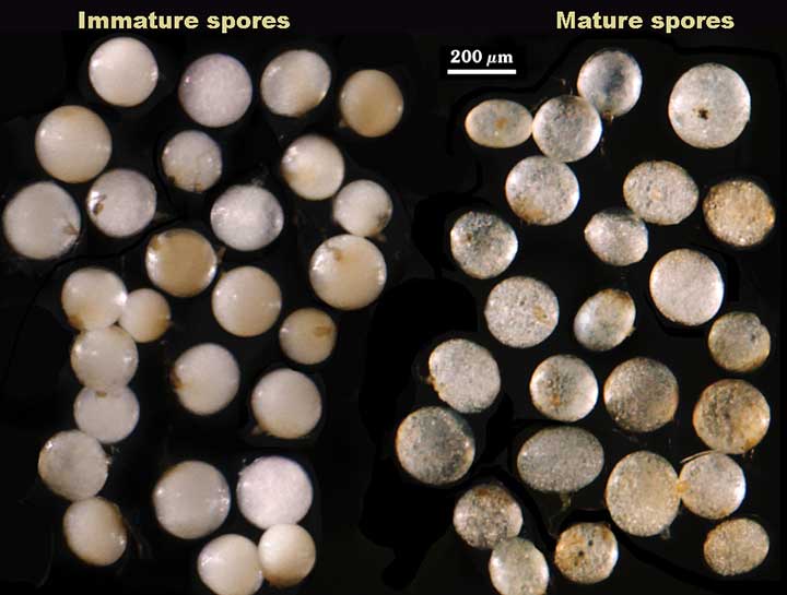
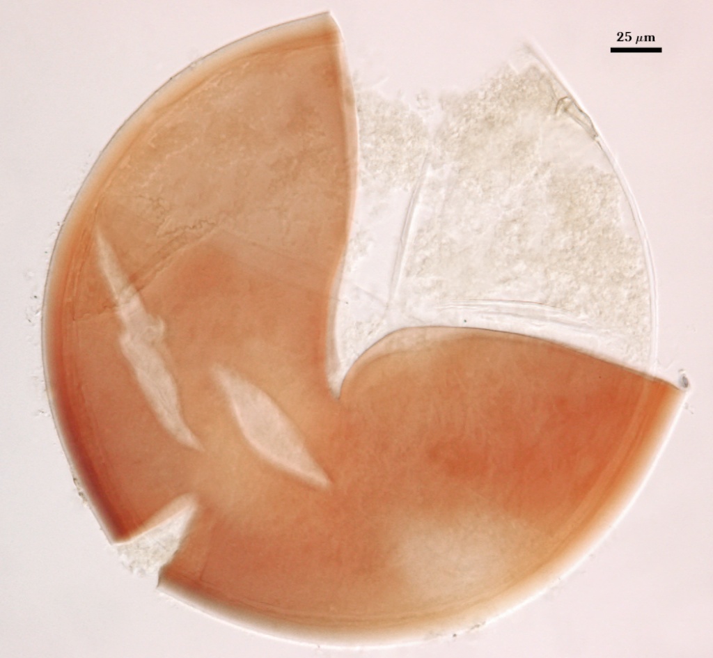
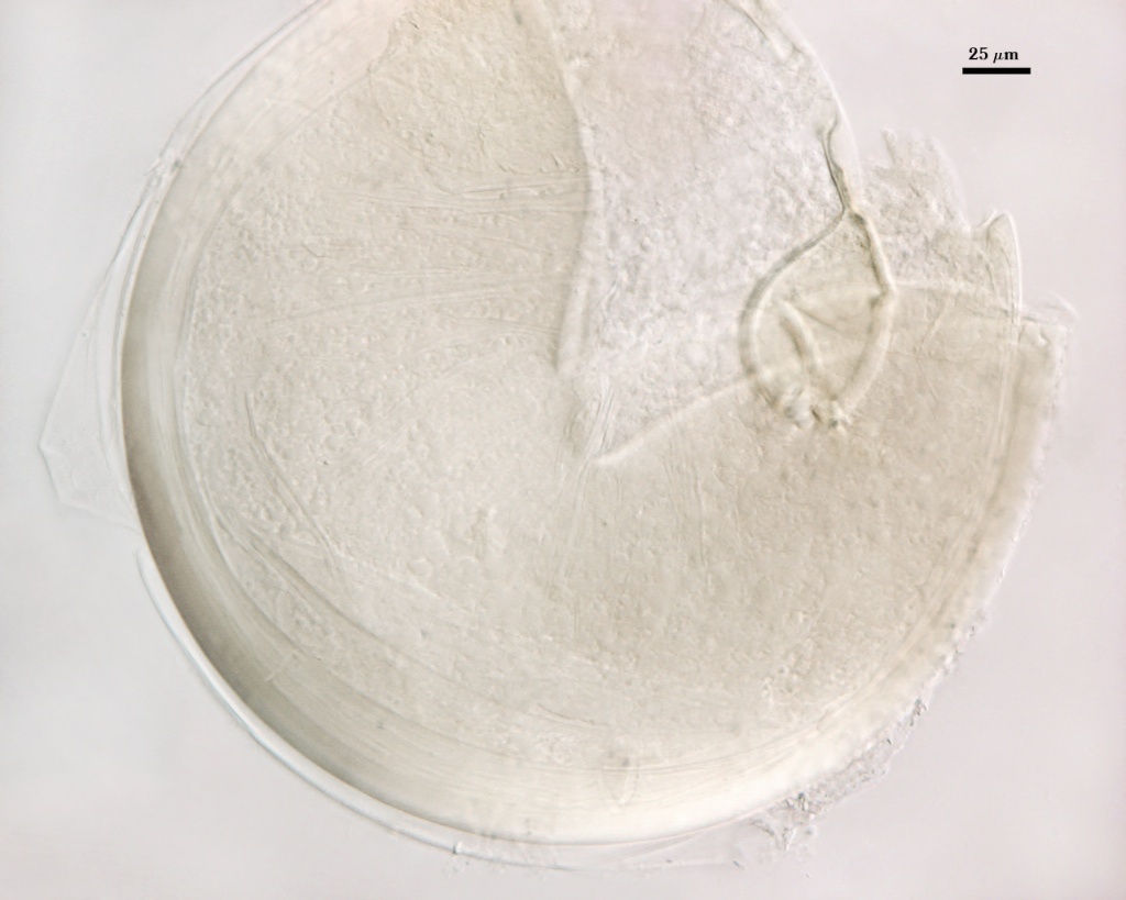
COLOR: Completely colorless (hyaline) to pale yellow.
SHAPE: Globose to subglobose, sometimes ovoid.
SIZE DISTRIBUTION: 120-220 µm, mean = 162 µm (n = 80). Within the same range as the wild-type strains.
Subcellular Structure of Spores
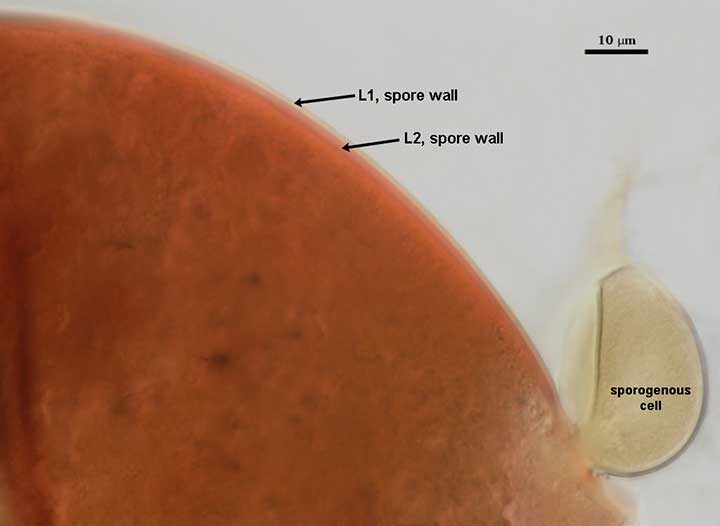
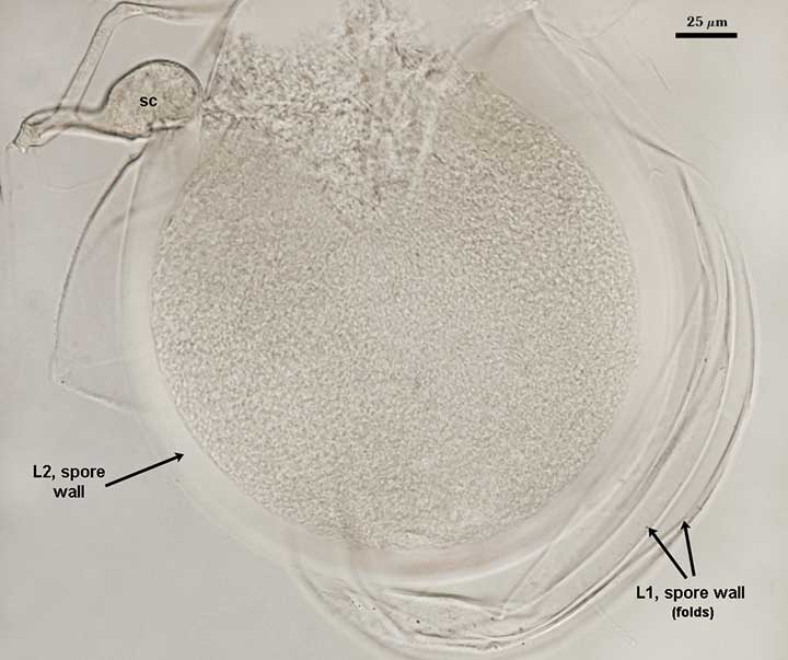
SPORE WALL: Three layers (L1, L2, and L3) with the middle layer (L2) undergoing a transformation identical to that observed in spores of wild-type strains, except that the transformation does not progress beyond the “amorphous” state. The sequence in the differentiation of this layer is chronicled in the photograph sequence below (from left to right). This mutant does not diverge significantly from wild-type strains using the LSU gene as a marker (Morton and Msiska, 2010). Therefore, the dramatic different in spore wall phenotype is a population-level variant that become fixed and stable. This mutant exemplifies a morphological difference that has no phylogenetic significance at the species level or above.
L1: An outer permanent rigid layer with tightly packed short rounded warts 1.0-2.5 µm high, pale brown (0-20-50-10) in color.
| Amorphus State | |||
|---|---|---|---|
 |  |  |  |
| Amorphus State (cont't) | |||
|---|---|---|---|
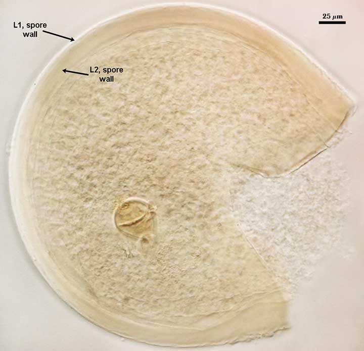 | 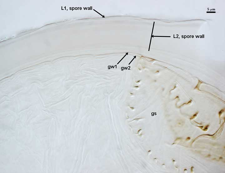 |  |  |
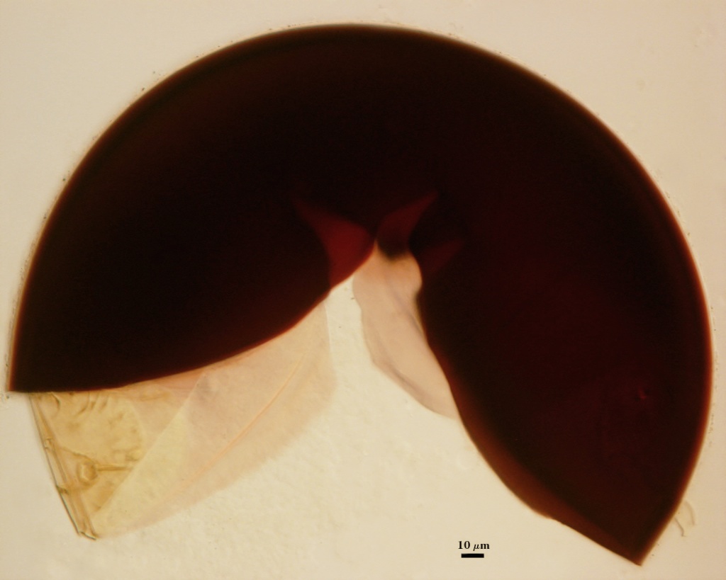
L2: A layer that has no sublayers, but is of a semi-pliable consistence (hence the term amorphous), hyaline to very pale yellow, thickness highly variable, from 10-32 µm. This layer stains dark red-purple to almost black in Melzer’s reagent. This reaction and the “amorphous” phenotype are correlated across all glomeromycotan species.
L3: Likely present, but cannot be detected because of wrinkling that occurs on the inner surface of L2 when pressure is applied.
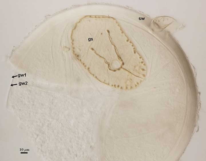
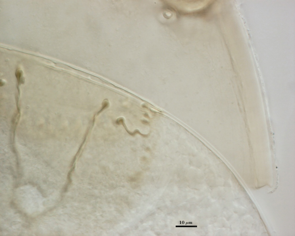
GERMINAL WALLS: Two bilayered flexible hyaline inner walls (gw1 and gw2) that are synthesized consecutively after the spore wall has completed differentiation. They are formed completely separate from the spore wall, in exactly the same way as in spores of wild type strains of D. heterogama.
GW1: No different from gw1 in spores of wild-type strains of D. heterogama
GW2: No different from gw2 in spores of wild-type strains of D. heterogama
Subtending Hypha
SPOROGENOUS CELL WALL STRUCTURE: Same as that of D. heterogama wild-type strains, except L2 does not become yellow-brown.
Germination Shield
Indistinguishable from that found in spores of the wild-type strains of D. heterogama. See images above at the end of the spore developmental sequence.
Auxiliary Cells
Indistinguishable from those produced by wild-type strains of D. heterogama.
Mycorrhizae
Indistinguishable from that shown for the wild-type strains of D. heterogama.
Notes
This mutant was started from a population of spores inoculated onto sorghum in 1991 and subcultured repeatedly since then at 1-2 year intervals of storage. Spores of the mutant are more susceptible to degradation in storage, and so they were cultured more often than wild-type strains. The albino phenotype has never reverted back to the wild-type, so it now has the same stability as all other traits used to define species.
The divergence in this albino mutant appears to be one more more mutations that prevented expression of events that transformed the “amorphous” L2 layer to the typical rigid laminae found in spores of the wild-type as well as any other species in Gigasporaceae. The absence of any perturbation to synthesis of the two germinal walls and the germination shield (identical to wild-type strains) provides the first empirical evidence that spore wall development occurs independently of inner wall synthesis.
The images below can be uploaded into your browser by clicking on the thumbnail or can be downloaded to your computer by clicking on the link below each image. Please do not use these images for other than personal use without expressed permission from INVAM.
High Resolution Images | |||
|---|---|---|---|
|  | ||
 |  | ||
 | | ||
Reference
- Morton, J. B. and Z. Msiska. 2010. Ontogeny and phylogeny of a Scutellospora heterogama mutant, with implications for species variation in Glomeromycota. Fungal Biology 114:410-420.