Rhizophagus irregularis
(reference accession PL112)
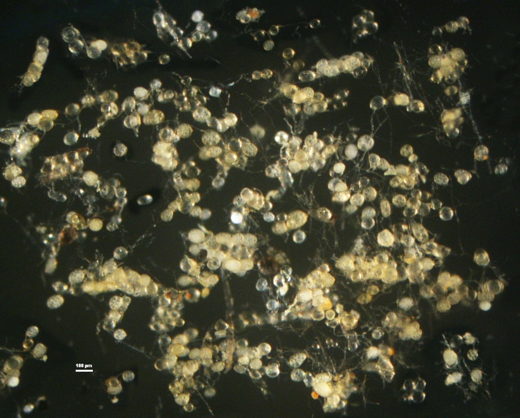
Whole Spores
COLOR: Almost hyaline (0-0-10-0) to yellow brown (0-10-40-0), but the range is highly variable even within spore populations of the two strains currently in the collection (PL112, DN201). Spores borne singly and in loose aggregates of a variable number of spores.
SHAPE: Globose, subglobose, ovoid, oblong, or irregular enough to sometimes appear knobby.
SIZE DISTRIBUTION: 70-165 µm in diameter (long axis).
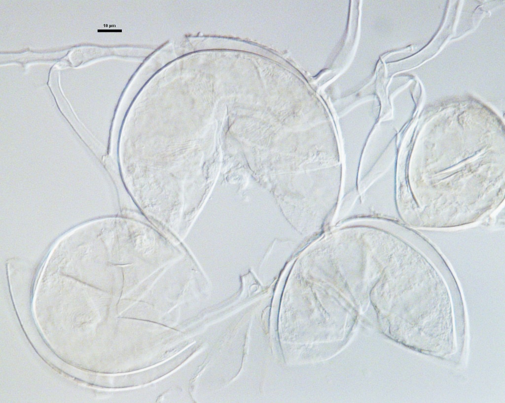
Subcellular Structure of Spores
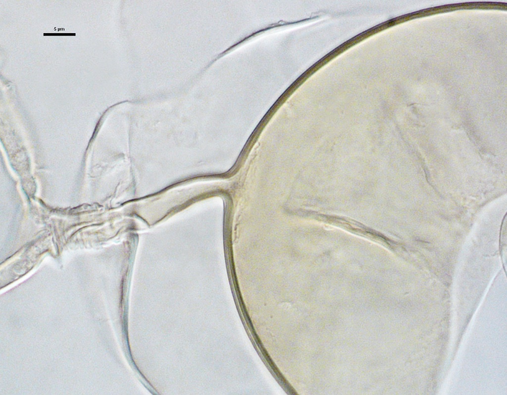
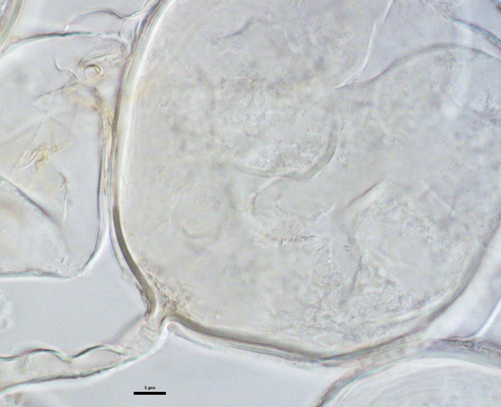
SPORE WALL: Three layers (L1, L2 and L3), with only the first layer present in juvenile spores and subtending hyphal wall (so they are colorless); L2 and L3 then formed sequentially in both the spore wall and in the wall of the subtending hypha.
L1: Outermost layer, hyaline, 0.5-1.5 µm thick, disintegrating at maturity with no reaction in Melzer’s reagent.
L2: Adherent to the outer layer (L1), hyaline, 1.0-4.9 µm thick (mean of 3.2 µm) when intact in young spores. With age, this layer degrades at a similar rate to L1 and appears granular or sloughs in patches. L1 and L2 are either absent or present as patchy remnants in mature spores.
L3: A permanent laminate layer, pale yellow (0-10-20-0) to yellow-brown (0-0-80-0 to 5-0-80-0) in transmitted light, 2.0-5.5 µm (mean = 3.9 µm) in mature spores. This layer stains a dark red-brown color in Melzer’s reagent.
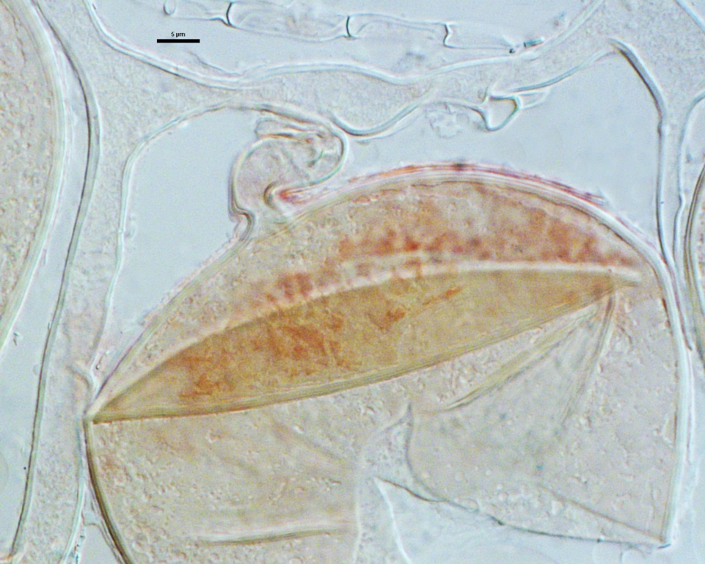
Subtending Hypha
COLOR: Similar to that of the L3 layer of the spore wall (pale yellow to yellow-brown)
SHAPE: Usually cylindrical to slightly flared.
WIDTH: 7.4-19 µm (mean = 11.2 µm).
COMPOSITE WALL THICKNESS: 2.0-7.6 µm.
WALL STRUCTURE: Three layers (L1, L2 and L3) that are continuous with the three layers of the spore wall. The two outer layers are the only ones present in early stages of spore formation, and both thin or degrade with spore maturation. Hyphae attached to spores can react lightly in Melzer’s reagent (a continuation of the L3 layer of the spore wall).
OCCLUSION: Recurved septum (from inner lamina of L3 spore wall layer), plug, or open.
Germination
A germ tube emerges from the lumen of the subtending hypha. It appears to arise from the innermost sublayer of L3.
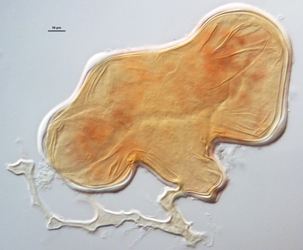
Mycorrhizae
Arbuscules and hyphae in cortical cells of sorghum and corn astain darkly in trypan blue. Numerous vesicles (or spores) often form near entry points along with the arbuscule-hyphal network.
Spores often form as dense clusters within roots, but also are patchily distributed singly or in clusters of only a few spores. There is a higher proportion of irregularly-shaped spores than observed with R. intraradices.
Notes
Both strains of R. irregularis were labeled as R. intraradices for a number of years. LSU sequence data tends to separate these strains from the many R. intraradices strains in the collection into a sister monophyletic clade. However, this single gene phylogeny may misrepresent the separation of these strains as a distinct species. The morphological differences are minor, many overlap and generally are within the realm of variation found in other glomoid species, including the much emphasized differential reactions in Melzer’s reagent (L1/L2 for R. intraradices and L3 for R. irregularis).
At issue is the perception that there may be other significant life history trait differences between these two “species” but all of the available evidence indicates otherwise. Mycorrhizal development is almost identical in both “species” with abundant vesicular colonization that seems to transition into formation of intraradical spores. Infectivity also is quite similar, with intraradical spores, mycorrhizal roots and external hyphae equally highly infective (maybe even moreso than spores).
The images below can be uploaded into your browser by clicking on the thumbnail or can be downloaded to your computer by clicking on the link below each image. Please do not use these images for other than personal use without expressed permission from INVAM.
High Resolution Images | |
|---|---|
 |  |
 |  |
 |  |
Links to Gene Sequences in Genbank
Reference
- Schenck, N.C.; Smith, G.S. 1982: Additional new and unreported species of mycorrhizal fungi (Endogonaceae) from Florida. Mycologia 74(1): 77-92.