Acaulospora kentinensis
(reference accession TW103)
= Entrophospora kentinensis
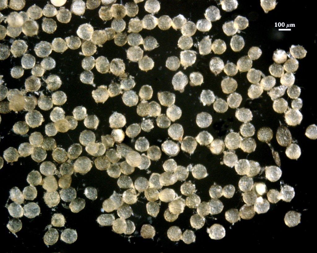
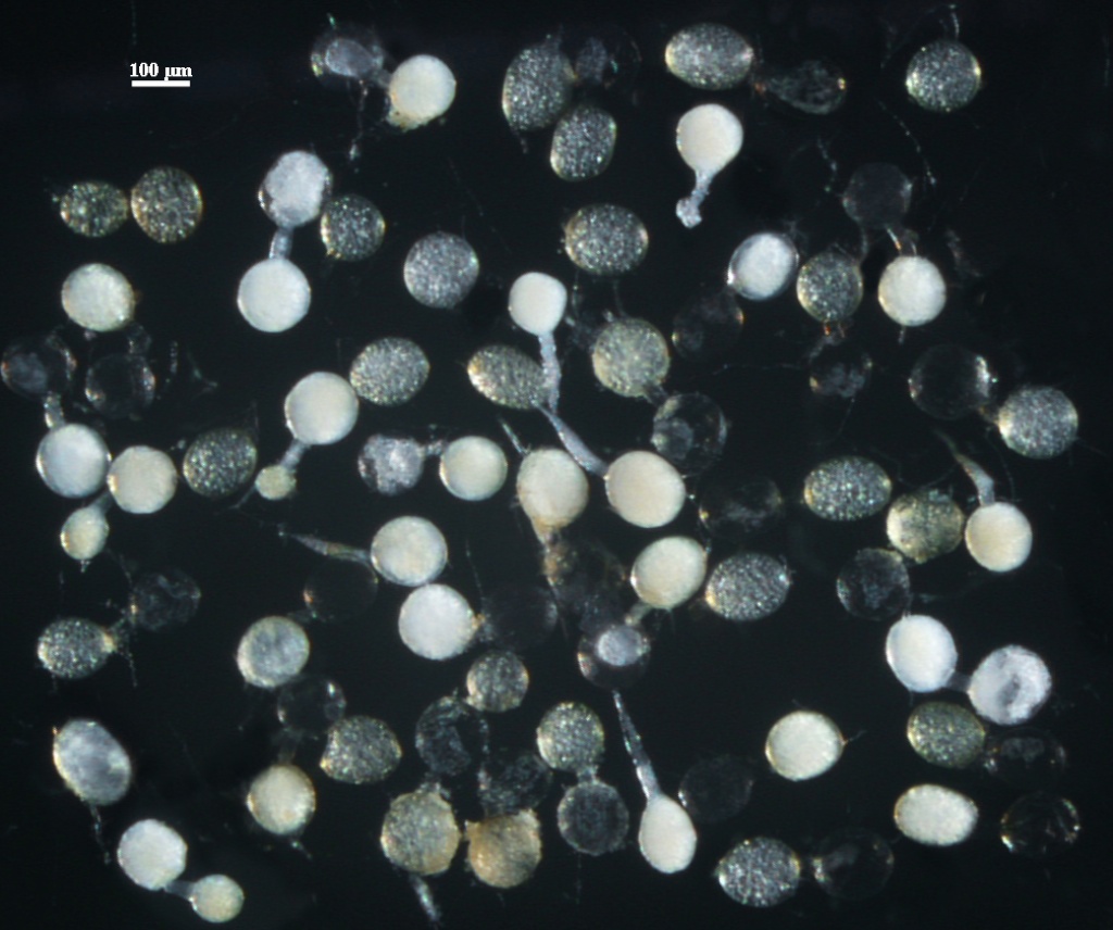
Whole Spores
| Spore Growth | |
|---|---|
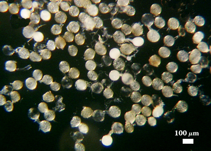 | 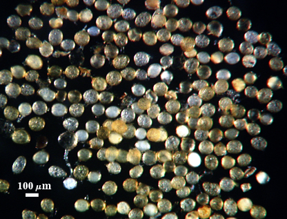 |
 COLOR: Orange-brown (0-20-60-0) to dark orange-brown (0-10-60-0), with many between these two extremes (0-40-100-0). Contents of juvenile spores are dense, so spores appear much more opaque than at maturity.
SHAPE: Globose to subglobose, more rarely ellipsoid or irregular.
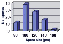
SIZE DISTRIBUTION: 80-160 µm, mean = 109 µm (n = 102).
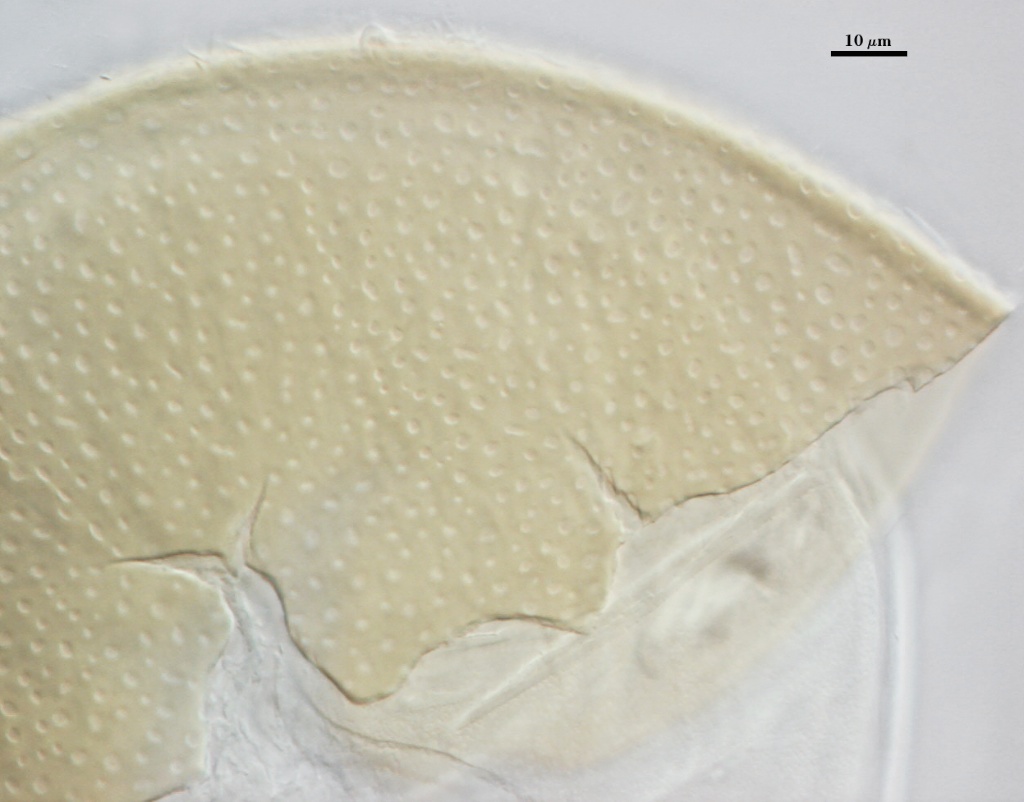
Subcellular Structure of Spores
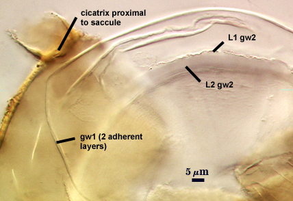
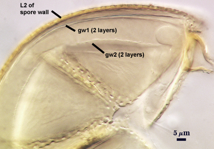
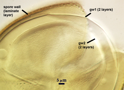
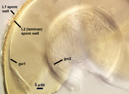
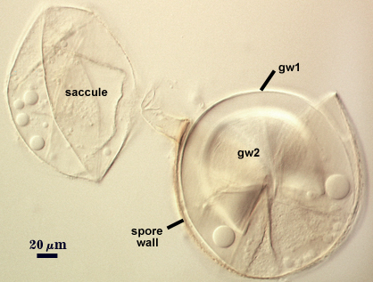
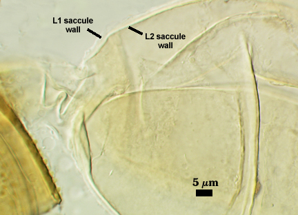
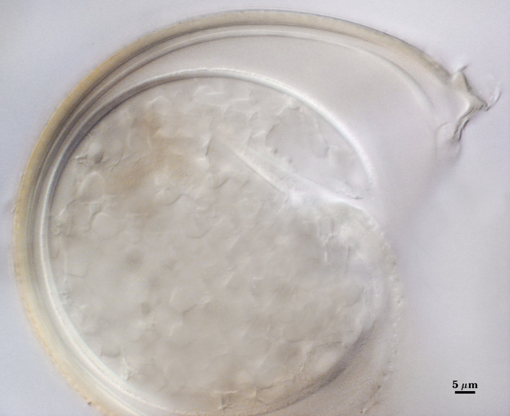
SPORE WALL: Two layers (L1 and L2), the outer layer continuous with the wall of the neck of the parent sporiferous saccule and sloughing; the inner layer becoming the spore wall, rigid, and ornamented.
L1: A hyaline layer that is continuous with the wall of the saccule neck; 0.5-0.8 µm thick; usually present on spores with saccules attached and sloughed when saccules become detached or collapse and degrade.
L2: A layer consisting of yellow-brown (0-30-100-0) to darker yellow-brown (0-30-100-10) sublayers (or laminae) that is seamlessly encloses the spore contents. Thickness ranges from 2.4-4.4 µm thick (mean of 3.3 µm). At maturity, the pore between spore and saccule neck is closed by continuous sublayers of this layer, with remnants of the saccule neck appearing as a ridge and leaves a wide scar (or cicatrix) on the spore surface.
| Spores mounted in PVLG | |||
|---|---|---|---|
|
|
|
|
| Spores in PVLG & Melzer’s reagent | |
|---|---|
|
|
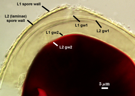
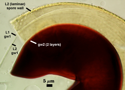
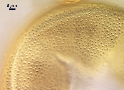
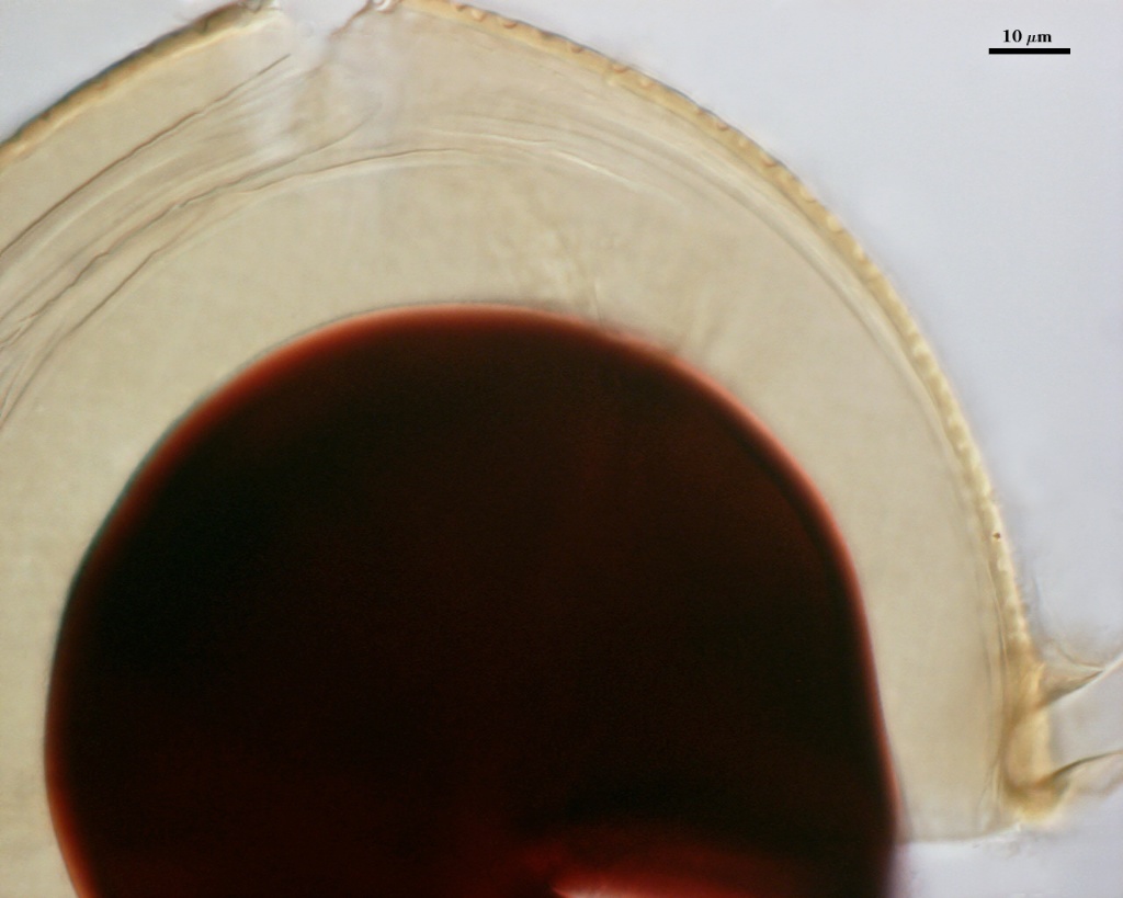
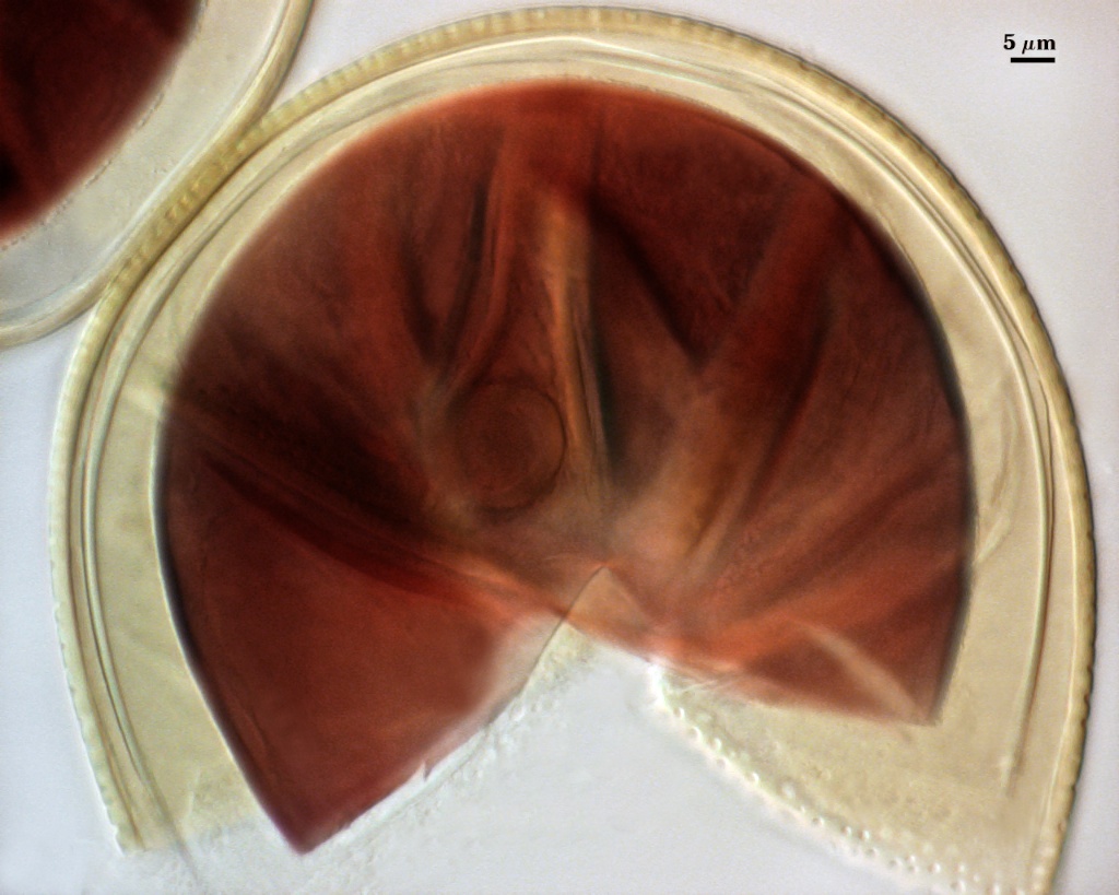
GERMINAL WALLS: Two flexible hyaline inner walls (gw1 and gw2) are readily seen in broken spores.
GW1: Often gives the appearance of being a single layer because the two layers present tend to be adherent; near equal thickness; together1-2 µm thick.
GW2: Consisting of two layers that also usually are adherent. L1 is 0.5-0.8 µm thick, with granular excrescence (or “beads”) that tends to become dislodged and float away with applied pressure. These “beads” are stabilized after preservation in formalin, but otherwise may be absent on mounted spores within a few months of storage. L2 is plastic enough that it has been termed “amorphous”. It can range in thickness from 4-12 µm in PVLG-based mountants, depending on amount of pressure applied to it while breaking the spore; staining red-purple (20-80-20-0) to dark red-purple (40-80-60-0) in Melzer’s reagent. This layer is nonreactive in youth and gradually becomes more reactive as differentiation is completed.
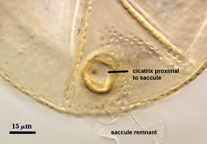
Cicatrix
Two are evident on spores, one proximal to the sporiferous saccule and one on the opposite side of the spore. The former is at least twice the diameter of the latter. 1 µm (mean = 21.7 µm) for the cicatrix proximal to the saccule, and 6-15 µm (mean = 10.8 µm) for the cicatrix distal to the saccule.
| Cicatrices | |
|---|---|
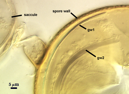 |  |
Sporiferous Saccule
COLOR: Salmon colored in youth, gradually becoming white and then hyaline as the contents are hydrolyzed or disintegrate.
SHAPE: Subglobose to slightly oblong.
SIZE: 100-110 µm, mean = 102.4 µm.
SACCULE WALL: One hyaline layer, 1.0-1.4 µm; surface smooth.
DISTANCE FROM SACCULE TO SPORE: 20-70 µm.
| Separated Saccule | |
|---|---|
 |  |
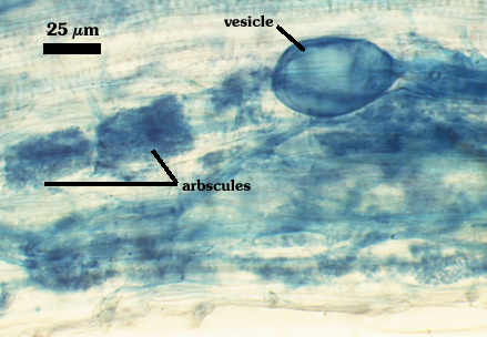
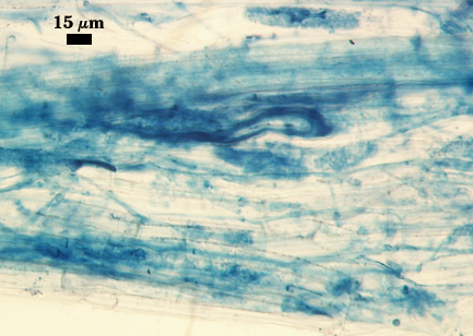
Mycorrhizae
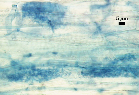
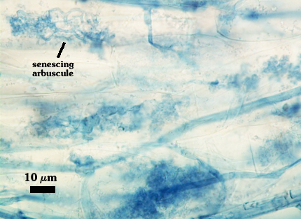
Arbuscules generally stain lightly in trypan blue, making observation of fine architecture difficult. Intraradical hyphae and vesicles stain with much more varying intensity, from almost invisible to quite dark. Infection units appear to be patchily distributed in red clover, sudangrass, and corn, with oblong to irregular vesicles often forming in localized clusters.
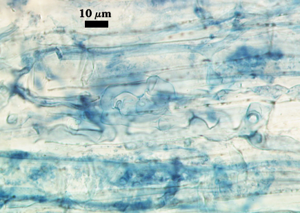
| Arbuscules in cortical cells of corn | |||
|---|---|---|---|
 |  | 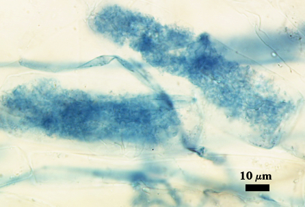 | 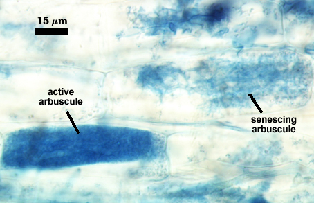 |
| Mycorrhizae in corn (emphasizing distribution of infection units and vesicles) | |||
|---|---|---|---|
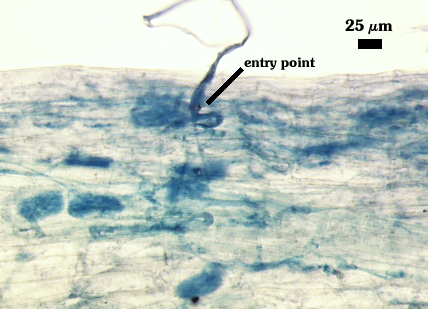 |  |  |  |
Germination
Not reported by Wu and Liu (1995) and not observed in pot cultured spores to date. It is assumed that a germ tube arises from a hyaline “germination orb” that forms on the innermost germinal wall (gw2).
Notes
So far, this species has been found only in Taiwan and Cuba. It has been cultured in standard potting mixes as well as in aeroponic chambers (Wu, personal communication). This species is somewhat unique in the genus in the robustness of the saccule neck wall, so that saccules often remain attached or when detached, a highly visible proximal cicatrix remains.
Wu and Liu (1995) do not report the granular nature of the surface of the outer layer of the innermost germinal wall (gw2). This is significant, because it appears to be a highly conserved trait shared by all species of Acaulosporaceae.
The images below can be uploaded into your browser by clicking on the thumbnail or can be downloaded to your computer by clicking on the link below each image. Please do not use these images for other than personal use without expressed permission from INVAM.
High Resolution Images | |
|---|---|
 |  |
 |  |
 |  |
 | |
Reference
- Wu, C.-G. and Y.-S. Liu. 1995. Glomales of Taiwan: V. Glomus chimonobambusae and Entrophospora kentinensis, spp. nov. Mycotaxon 53:283-294.