Acaulospora lacunosa
(reference accession WV201)
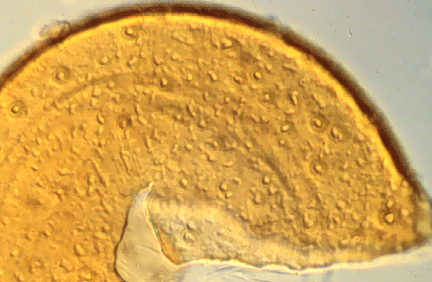
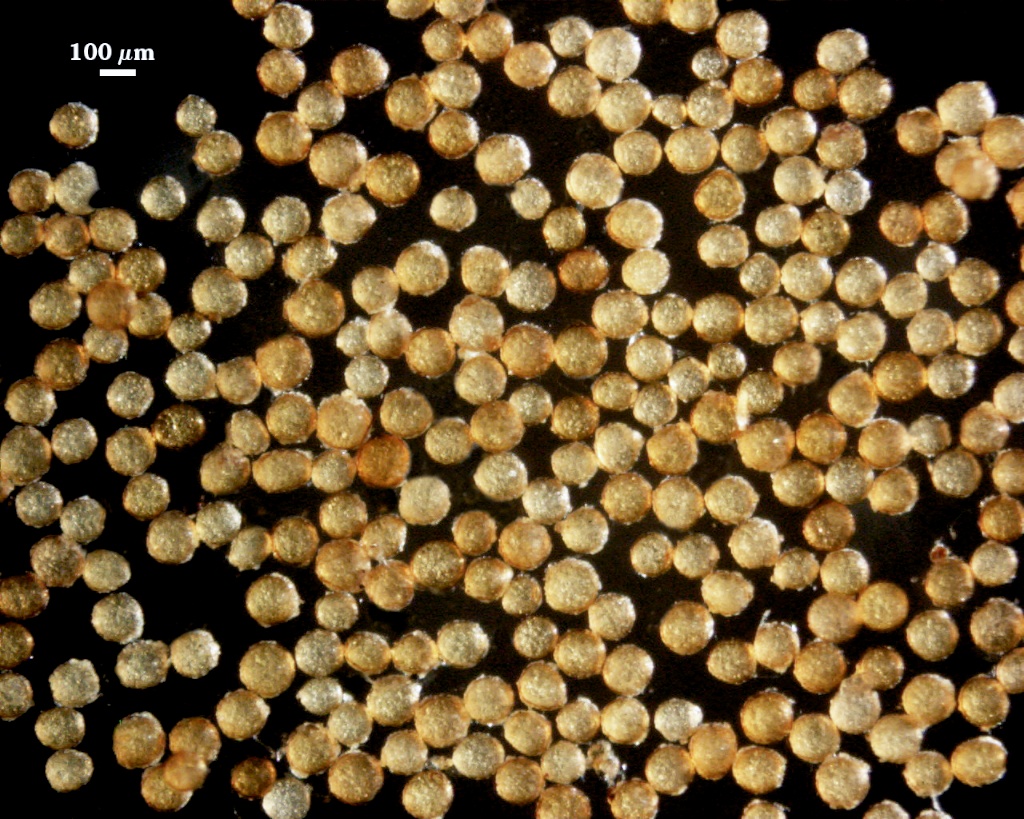
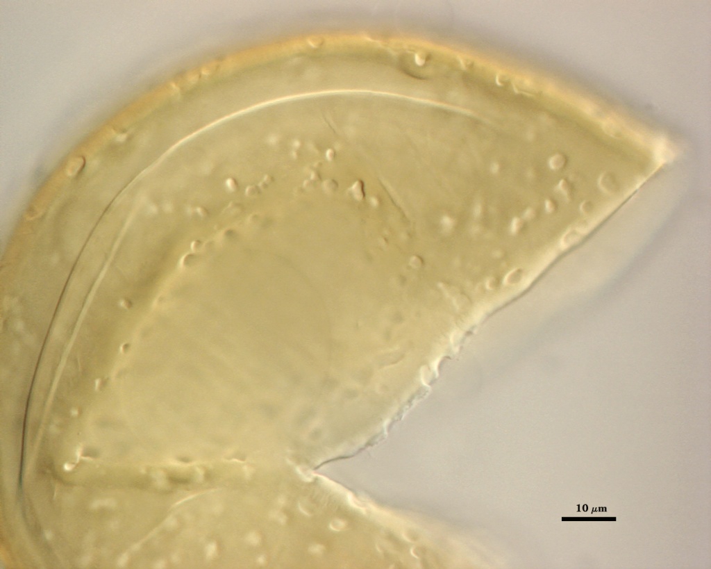
Whole Spores
| Spherical Saccules | |
|---|---|
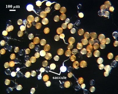 | 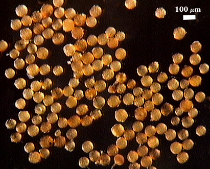 |
 COLOR: Orange-brown (0-40-80-0) to dark orange-brown (0-60-100-10),
SHAPE: Mostly globose, subglobose, ocasionally ellipsoid
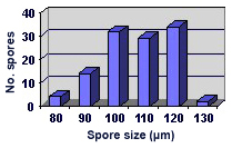
SIZE DISTRIBUTION: 80-130 µm, mean = 120 µm (n = 142)
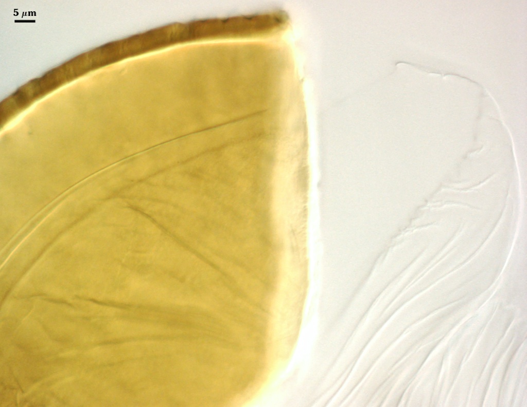
Subcellular Structure of Spores
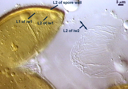
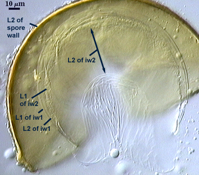
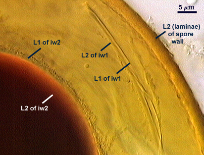
SPORE WALL: Two layers (L1, L2); the outer layer continous with the wall of the neck of the parent sporiferous saccule and the inner layer synthesized with development of the spore.
| Spore wall with irregular dentations | |
|---|---|
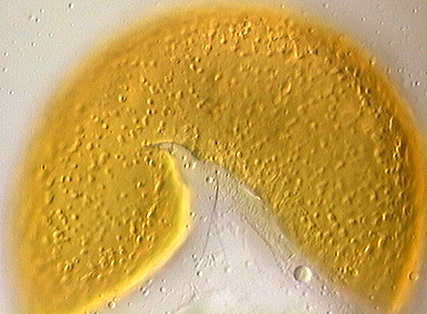 |  |
L1: A hyaline layer that is continuous with the wall of the saccule neck; 0.5-0.8 µm thick; usually present on spores with saccules attached and sloughed when saccules become detached or collapse and degrade.
L2: A layer consisting of yellow-brown (0-30-100-0) to darker yellow-brown (0-30-100-10) sublayers (or laminae) that is seamlessly encloses the spore contents. Thickness ranges from 2.4-4.4 µm thick (mean of 3.3 µm). At maturity, the pore between spore and saccule neck is closed by continuous sublayers of this layer, with remnants of the saccule neck appearing as a ridge and leaves a wide scar (or cicatrix) on the spore surface.
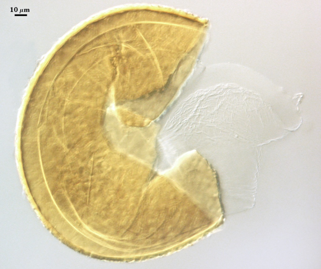
| In PVLG | |
|---|---|
|
|
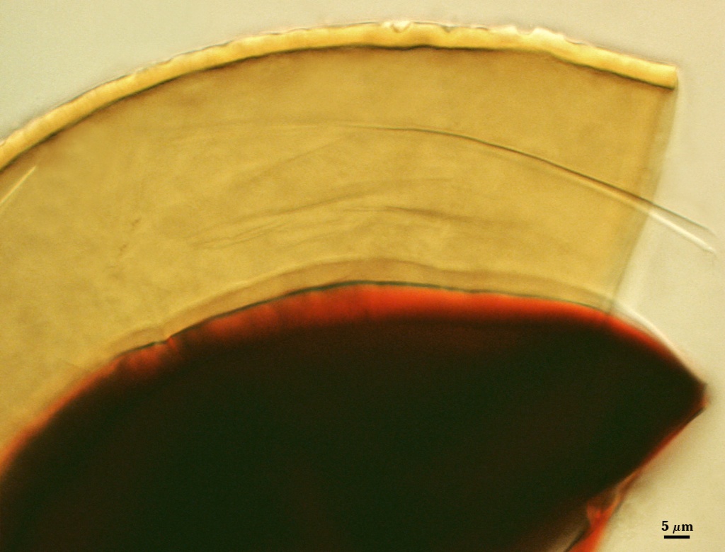
| Spores in PVLG & Melzer’s reagent | |
|---|---|
|
|
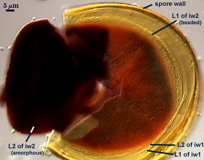
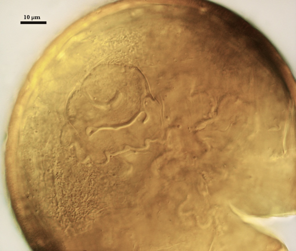
FLEXIBLE GERMINAL WALLS: Two walls (gw1 and gw2) are present that readily separate from each other and from the spore wall.
GW1: A bilayered hyaline flexible wall that separates easily from the spore wall and thus can be seen readily. Layers separate somewhat in most crushed spores. Both layers is of near equivalent thickness, 0.5-1.3 µm.
GW2: Consisting of two adherent hyaline layers. L1 is 0.5-1.0 µm thick (measurable only in PVLG), with granular excresences (or “beads”) that tend to become dislodged and float away with applied pressure. These “beads” are stabilized after preservation in formalin, but otherwise may be absent on mounted spores within a few months of storage. L2 is plastic enough that it has been termed “amorphous” (originally defined as an “amorphous wall”). It is 1-8 µm thick in PVLG-based mountants, depending on amount of pressure applied to it while breaking the spore; staining red-purple (20-80-20-0) to dark red-purple (40-80-60-0) in Melzer’s reagent. After fixation or in water, this layer loses its plasticity and is 1.5-4 µm thick.
Cicatrix
Scar indicating region of contact between spore and saccule neck during spore synthesis; very narrow lip (not measured) circumscribing the scar, circular to oval-shaped, 5-7.2 µm at widest.
Sporiferous Saccule
COLOR: Hyaline (see photo of whole spores at top of page).
SHAPE: Mostly globose to subglobose, occasionally irregular.
SIZE: 100-128 µm
SACCULE WALL: One layer, smooth surface, 0.8-1.2 µm thick
DISTANCE FROM SACCULE TO SPORE: 40-90 µm.
Germination
An ovoid “germination orb” forms on gw2, from which germ tubes form and penetrate through the spore wall. This orb is difficult to see except in older spores where contents have cleared with fusion of lipid globules in the spore lumen.
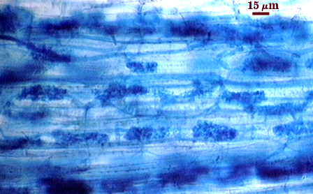
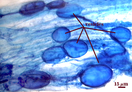
Mycorrhizae
Arbuscules and intraradical hyphae stain faintly in trypan blue, although intensity can be highly variable with age of the mycorrhizae and host plant. Infection units appear to be patchily distributed with oblong to irregular vesicles often forming in small clusters.
| All mycorrhizal structures in corn roots. | |
|---|---|
 |  |
Notes
In the protologue, the size range of this species is reported as 98-182 µm, but this included field collected as well as cultured specimens.
High Resolution Images | |
|---|---|
 |  |
 |  |
 |  |
Reference
- Morton, J. B. 1986. Effects of mountants and fixatives on wall structure and Melzer’s reaction in spores of two Acaulospora species (Endogonaceae). Mycologia 78:787-794.