Gigaspora margarita
(reference accession WV205A)
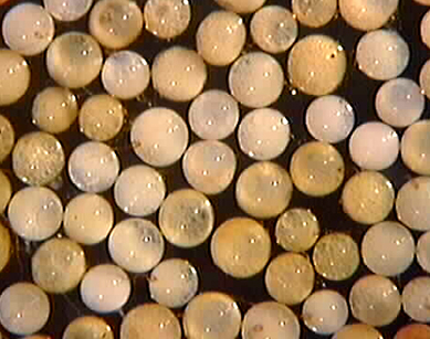
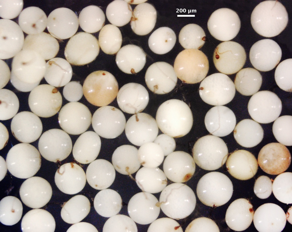
Whole Spores
 COLOR: White to cream (0-5-30-0) in many spores, darker yellow (0-10-40-0) in some generations or some isolates.
SHAPE: Globose to subglobose.
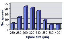
SIZE DISTRIBUTION: 260-400 µm, mean = 321 µm (n = 98).
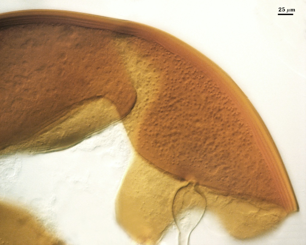
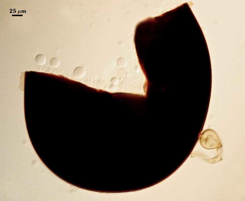
Subcellular Structure of Spores
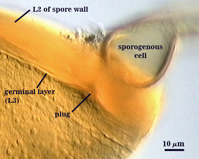
SPORE WALL: Three layers (L1, L2, and L3), the first two adherent and of equal thickness in juvenile spores, with L2 thickening as the spore wall is differentiated and L3 differentiating as a prelude to germ tube formation.
| In PVLG | In PVLG + Melzer's Reagent |
|---|---|
|
|
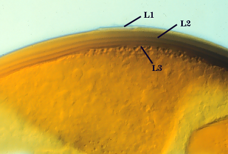
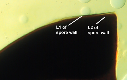
L1: An outer permanent rigid layer, smooth, adherent to inner laminae, pale brownish-yellow (0-10-30-0), 1.6-2.4 µm thick.
L2: A layer consisting of hyaline sublayers (or laminae) that increase in number with thickness are rigid, exhibit with some plasticity (swelling and spreading) when broken, yellow (0-10-80-0) to brownish yellow (0-10-100-0) in PVLG; 13-31 µm thick (mean of 22 µm). Thickness varies considerably even within one spore (14-23 µm); staining dark red-brown (20-80-100-0) to very dark red-purple (60-80-70-10) in Melzer’s reagent. In younger spores, sublayers merge and resemble “waves” without sharp transitions from ridge to trough. The extent to which this property is evident can be directly correlated with intensity and color of the reaction to Melzer’s reagent.
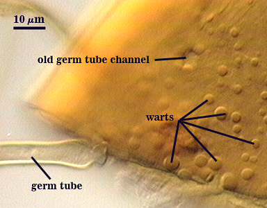
L3: A “germinal” layer that is concolorous and adherent with the laminate layer. This layer often is distinct only at the ultrastructural level, where it appears electron dense. Numerous “warts” or “papillae” form on the inner surface of this layer, and they are especially concentrated in regions where germ tubes form (usually in close proximity to the suspensor cell); warts 1.2-5 µm high in germinating spores and 2.5-3 µm wide.
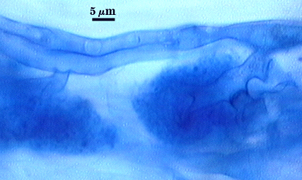
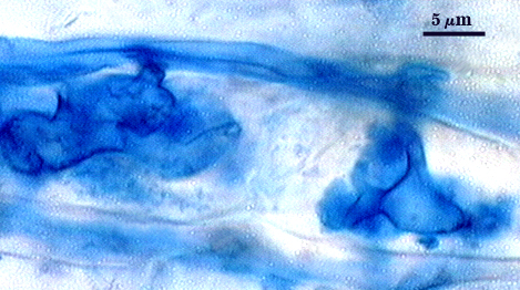
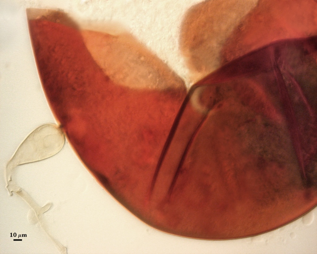
Subtending Hypha
WIDTH OF SPOROGENOUS CELL: 34-47 µm (mean = 42 µm)
SPOROGENOUS CELL WALL: Two hyaline layers (L1 and L2) probably are present (continuous with the first two layers of the spore wall), but only L2 is readily discernible at the level of the compound microscope.
L2: Brownish yellow (0-10-60-0), 3.2-6.4 µm thick near the spore and then thinning to 1.2-1.6 µm beyond the sporogenous cell.
OCCLUSION: Closure by a plug concolorous with the laminate layer of the spore wall.
Germination
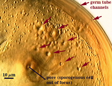
Germ tube forms in vicinity of warty protruberances on inner surface of L3 of the spore wall, hole through spore wall 6-8 µm wide, with the germ tube expanding immediately after emergence from the spore wall (12-15 µm wide).
| Germ tube emerging from the from spore wall | Eight holes where germination had occurred |
|---|---|
|
|
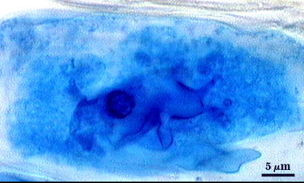
Auxiliary Cells
Cells in aggregates of 4-20, subglobose to ovoid to clavate, borne on tightly coiled hyaline hyphae, thin-walled (< 1 µm thick), hyaline to pale cream (0-0-10-0); each cell with narrow projections 1.5-2.0 µm wide and 2.0-10.0 µm high.
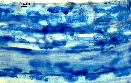
Mycorrhizae
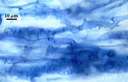
Intraradical arbuscules and hyphae consistently stain darkly in roots treated with trypan blue. Arbuscules produce fine-branches from a swollen basal hypha that is easiest to observe once tips begin to degrade. Intraradical hyphae 3-9 µm in diameter, with inflated areas up to 16 µm and knob-like projections distributed along length; usually densely coiled near entry points an in many cortical cells.
| Arbuscules in corn root cortical cells | ||
|---|---|---|
|
|
|
| Mycorrhizae in corn roots | |
|---|---|
|
|
Notes
Immature spores are salmon colored with a slight pink tint (0-10-20-0 to 0-20-60-0). The two layers of the spore wall are near-equivalent thicknesses before laminae develop from the inner layer (1.6-2 µm), with the outer layer not changing during differentiation of L2. Gigaspora ramisporophora was merged with Gi. margarita (Bentivenga and Morton, 1995) when cultures clearly identified as the latter species also produce pale yellow spores fitting the description of the former species. Both size and color can separate this species from Gi. rosea if the spores are examined shortly after extraction.
The images below can be uploaded into your browser by clicking on the thumbnail or can be downloaded to your computer by clicking on the link below each image. Please do not use these images for other than personal use without expressed permission from INVAM.
High Resolution Images | |
|---|---|
 |  |
 |  |
Reference
- Bentivenga, S. P. and J. B. Morton. 1995. A monograph of the genus Gigaspora incorporating developmental patterns of morphological characters. Mycologia 87: 720-732.