Glomus multicaule
Voucher Specimen
This description is a combination of information obtained from the protologue (Gerdemann and Bakshi, 1976), type specimens, and universal patterns of morphological organization and structure in Glomaceae. A living culture of this species has never been obtained by INVAM (or elsewhere that we know of).
Spores formed only singly in soil; ellipsoid, broadly ellipsoidal, subglobose, or occasionally triangular; 149-249×124-162 µm in diameter.
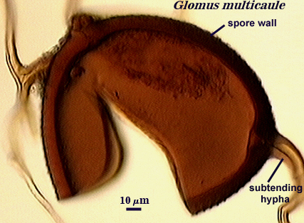
Spore wall described as having a single layer 8.6-34 µm thick (thickest at base of spore), with rounded projections 1.2-3.7 µm long regularly distributed over the spore surface.
Subtending hyphae, number of which varies from 1-4, with much of the material comprising type specimens having at least two hyphae rarely positioned adjacent to each other.
The multiple attached hyphae and very thick ornamented spore wall distinguish this species from other glomeromycotan fungi described to date.
(Note: Only thumbnail images were available, for spore image examples #2 & #3) | ||
|---|---|---|
 |  |  |
Reference
- Gerdemann, J. W. and B. K. Bakshi. 1976. Endogonaceae of India: Two new species. Trans. Br. Mycol. Soc. 66:340-343.