Dentiscutata nigra
(reference accession NC182)
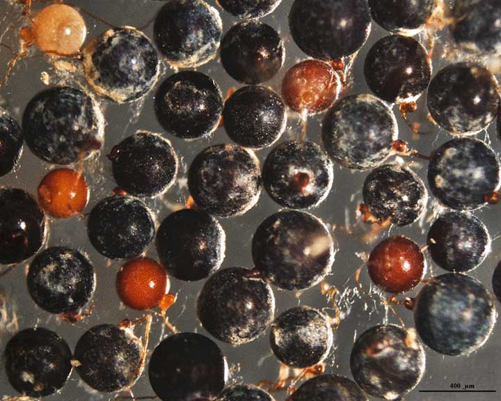
Whole Spores
(Relatively new culture, more information to come soon)
COLOR: Dark red-brown (40-80-100-10) to black. Immature spores are cream to pale yellow-brown, smooth surface, and dense contents under a dissecting microscope and in water.
SHAPE: Mostly globose.
SIZE DISTRIBUTION: 240-520 µm.
Subcellular Structure of Spores
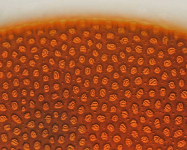
SPORE WALL: Two layers, the outer being smooth and the inner layer with a complex ornamentation pattern.
| Spore wall reticulation | Spore wall evenly spaced bumps inner wall |
|---|---|
|
|
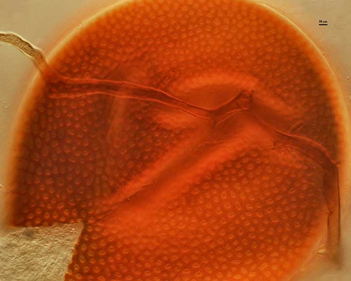
L1: Thin (2-3 µm), tightly adherent to L2. Indeterminant color because it can’t be viewed separately from L2, but both it and L2 are hyaline in youth.
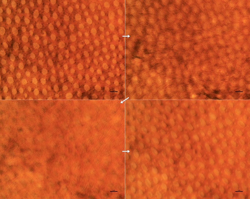
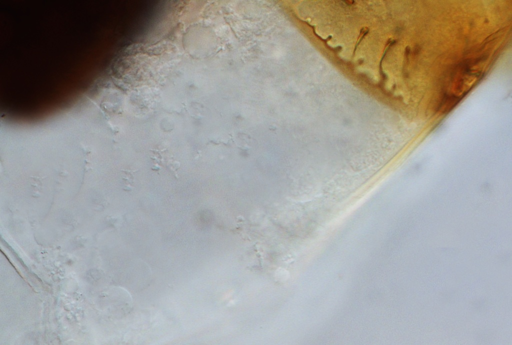
L2: Dark red-brown to almost black, 6-10 µm thick. with circular to oblong rings on the upper and lower surfaces with “wavy” spines in the center matrix that appear as a semi-circle in the center of each ring when viewed from above. Both of these layers form very quickly and are present in young spores that have yet to form any germinal walls within.
| Immature spore layers | |
|---|---|
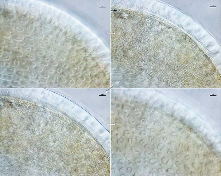 |  |
Above are two series of images of pale cream-colored immature spores mounted in PVLG; showing levels in L2 of the spore wall in a sequence from top to bottom surfaces (left to right, following the arrows). | |
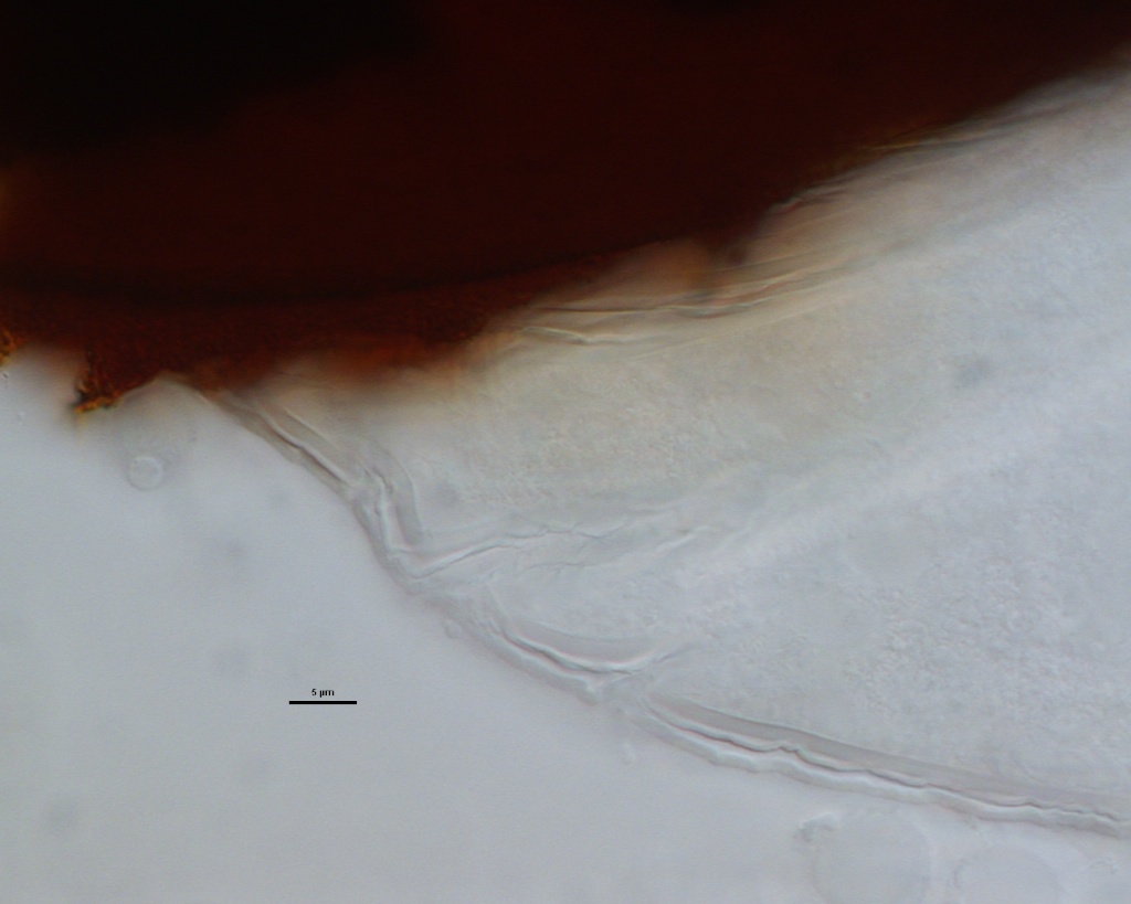
GERMINAL WALLS: Two bilayered flexible hyaline inner walls (gw1 and gw2) that are synthesized consecutively after the spore wall has completed differentiation. They are formed after the spore wall as completely differentiated. In many mature spores, both germinal walls tend to stay closely paired, even with applied pressure.
| GW1 GW2 thin lines | |
|---|---|
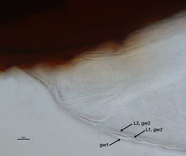 | 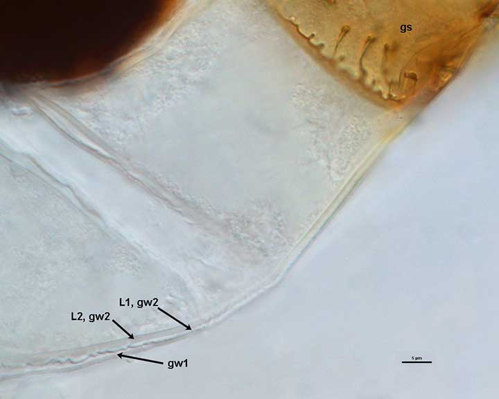 |
GW1: Two layers are formed (L1 and L2) that are tightly adherent. L1 is less than 0.5 µm thick; L2 is slightly thicker (0.8-1.5 µm). Both layers are thin enough that when tightly adherent, they appear as one layer.
GW2: Two layers are formed (L1 and L2) that are tightly adherent. The two germinal walls are not easy to separate or distinguish except when a germination shield is formed, which is positioned between the two walls.
Subtending Hypha
WIDTH OF SPOROGENOUS CELL: 24-36 µm (mean = 31 µm)
SPOROGENOUS CELL WALL STRUCTURE: Dark red-brown in young spores (cream-colored), with the spore wall becoming concolorous as it matures.
OCCLUSION: Closure by a plug concolorous with L2 of the spore wall.
Germination
COLOR: Pale yellow-brown (0-30-70-10) to darker orange-brown (0-40-100-10).
SHAPE: Oblong, with length approximately 1.5 times that of the width. Margin of the shields is fairly smooth, with only a few folds and attendant paired germ holes. Position of the shield is on gw2.
The images below can be uploaded into your browser by clicking on the thumbnail or can be downloaded to your computer by clicking on the link below each image. Please do not use these images for other than personal use without expressed permission from INVAM.
High Resolution Images | |
|---|---|
|  |
 |  |