Glomus pansihalos
Voucher Specimen
This description is a combination of information obtained from the protologue (Koske and Halvorson, 1989), type specimens, and universal patterns of morphological organization and structure in Glomaceae.
| Sporocarps | ||
|---|---|---|
 |  |  |
Sporocarps, when formed, are loose clusters without a peridium, 15×12 x 7 mm in size, pale to dark brown, containing a mix of spores, hyphae, roots, and the medium in which the sporocarps were formed.
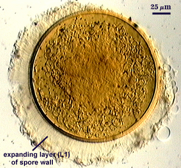
Spores globose, ellipsoid, to irregular in shape, 130-155×120-130 µm in diameter (with extremes of 108-180 µm), yellow-brown to dark brown-yellow in color.
Subcellular Structure of Spores
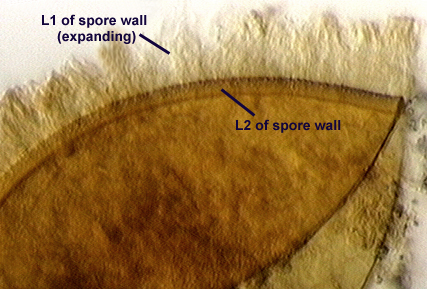
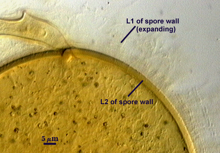
The spore wall is reported to consist of three layers (L1, L2, L3), but only L1-L2 were observed in any of the type material. It must be noted that all of the spores examined were either degraded or parasitized, so there was no good comparison with healthy specimens. Photographs in the protologue (Berch and Koske, 1986) suggest better specimens were examined by the authors. The outer layer (L1) is hyaline, swelling and expanding in lactic acid or PVLG and forming hyaline to pale yellow columns radiating outward from the spore surface as far as 100 µm. In water, this layer is rigid and 3-5 µm thick. The laminate layer (L2) is rigid, yellow-orange to brownish-orange, 3-8 µm thick, and reportedly covered with hemispherical 1×1 x 1 µm warts spaced apart by 1-5 µm. The inner most layer (L3) is yellow-orange, and 1-2 µm thick.
Subtending Hypha
The subtending hypha is single, straight or either flared or constricted, sometimes recurved (basically anything goes) at the point of attachment, pale yellow-brown in color, and 10-20 µm wide at the spore base. The hyphal wall consists of all layers of the spore wall, up to 5 µm thick, at the spore base, with the outer layer becoming dispersed with distance. The hyphal pore either is open or occluded by a septum formed by L3 of the spore wall (in the far right photo above?).
Reference
- Berch, S. M. and R. E. Koske. 1986. Glomus pansihalos: A new species in the Endogonaceae, Zygomycetes. Mycologia 78:832-836.