Cetraspora pellucida
(reference accession FL966)
= Scutellospora pellucida
Whole Spores
| Appearance of contents |
|---|
 |
| Accession FL966 | Accession NC118 |
|---|---|
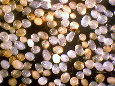 |  |
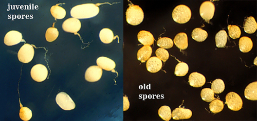
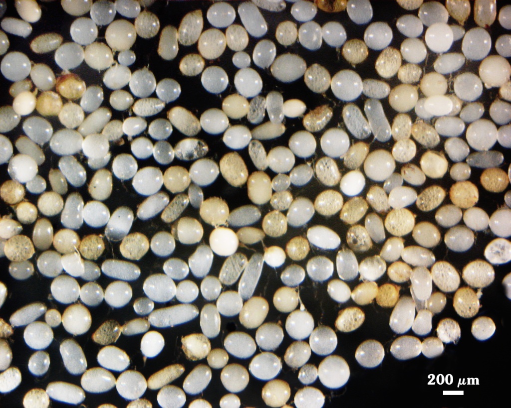
 COLOR: Hyaline/white in most recently formed spores to yellow-brown (0-5-40-0) in older spores (especially those from field soils).
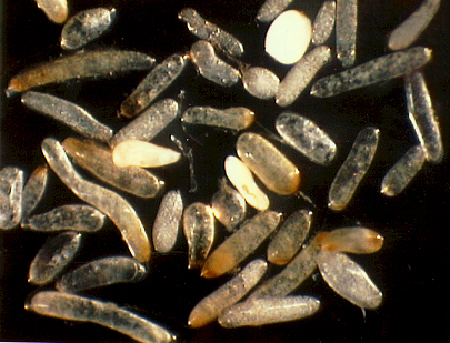
SHAPE: Globose, subglobose, often elliptical or strongly oblong.
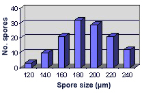
SIZE DISTRIBUTION: 120-240 µm, mean = 189 µm (n = 128).
Subcellular Structure of Spores
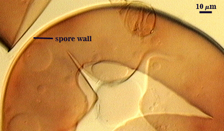
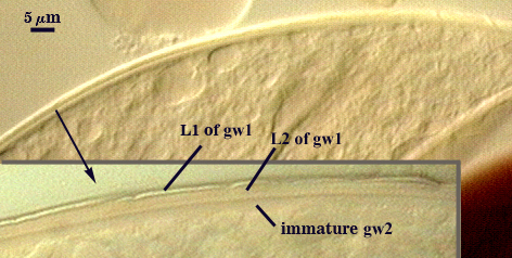
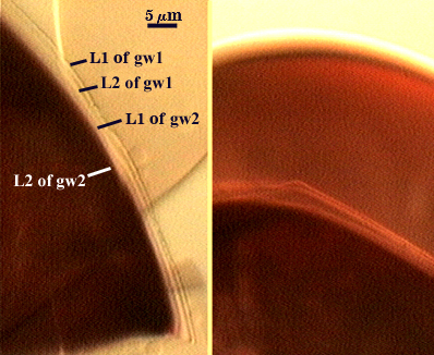
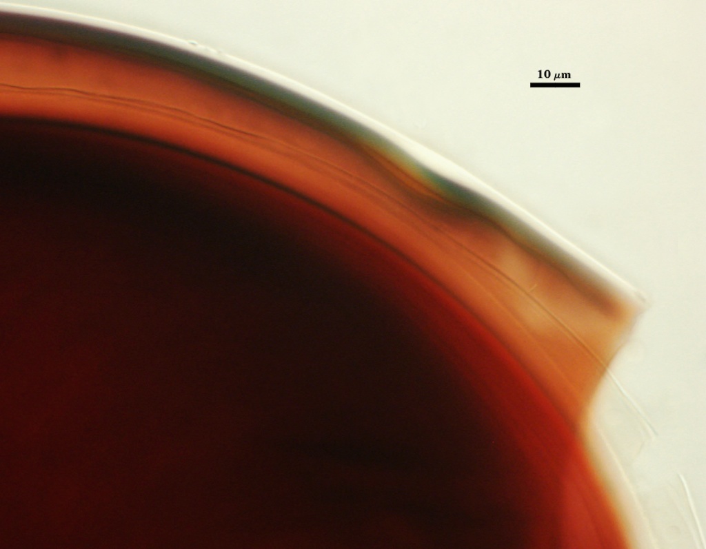
SPORE WALL: Three layers (L1, L2 and L3) that are adherent that in juvenile spores are of equal thickness, with the laminate layer (L2) thickening as the spore wall is differentiated. Below (left to right, click to enlarge) is a linear sequence in the differentiation of a spore wall and two flexible inner walls. It is uncertain when L3 appears developmentally.
| Spore wall | |||||
|---|---|---|---|---|---|
|
|
|
|
|
|
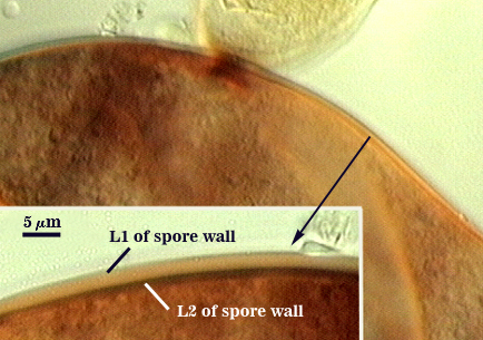
L1: An outer permanent rigid hyaline layer with a smooth surface, 1.8-5.0 µm thick and tightly adherent to L2. Easily distinguished from L2 when spores are placed in Melzer’s reagent, where L1 is nonreactive and L2 stains dark red-purple.
L2: A layer consisting of very fine adherent hyaline to pale yellow (0-0-20-0) sublayers (or laminae), 3.0-8.8 µm thick (mean of 6.5 µm) in mature spores; staining dark red-purple (40-80-40-0) to reddish-black (60-80-50-10) in Melzer’s reagent.
L3: A very thin hyaline flexible layer, < 1 µm thick, can be seen in vigorously crushed spores, usually only where it attaches to the spore wall near the occluding plug and the sporogenous cell region (not shown). A homologous layer was first detected in S. cerradensis and provided the impetus to search for a similar layer in other Scutellospora species.
| In PVLG | ||
|---|---|---|
 |  | |
| In PVLG & Melzer’s reagent | |
|---|---|
 | 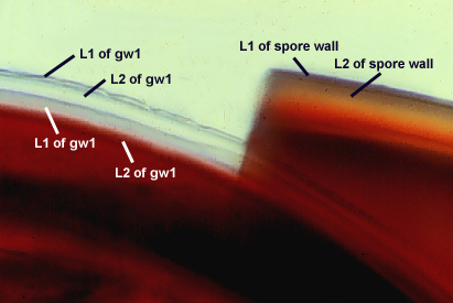 |
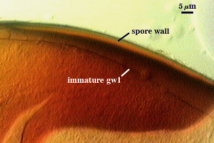
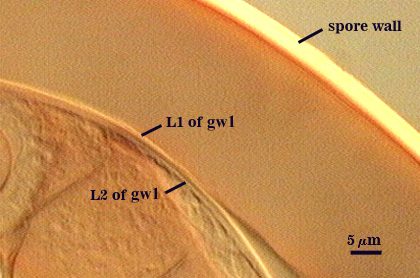
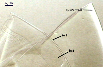
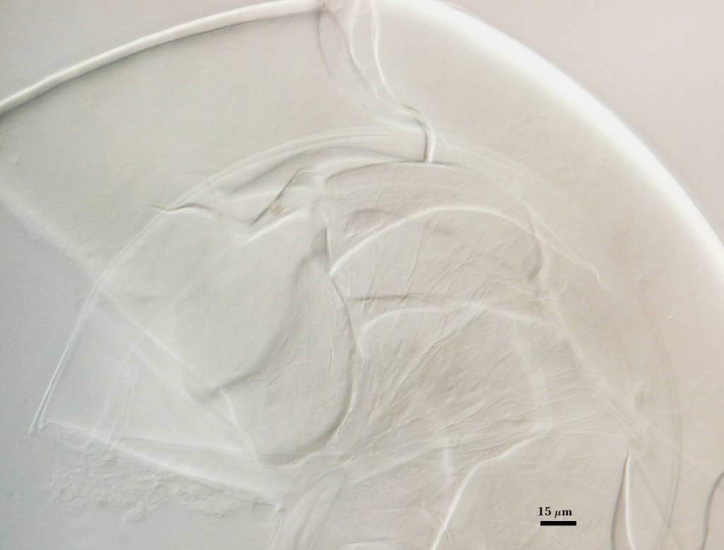
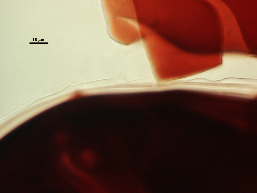
GERMINAL WALLS: Two bi-layered hyaline flexible inner walls (gw1 and gw2) are formed that readily separate from each other and from the spore wall. Both walls form in sequence and begin synthesis only after the spore wall has completed differentiation (see photos above)
GW1: Two layers (L1 and L2) that usually are adherent, but sometimes L1 separates slightly and produces numerous fine folds. L1 is less than 0.5 µm thick and L2 is 1.0-2.8 µm thick. Neither layer reacts in Melzer’s reagent.
GW2: Two layers (L1 and L2) that rarely separate. L1 is 2.0-4.8 µm thick and can produce a weak pink reaction (0-10-20-0) in Melzer’s reagent that is detected only when it separates from L2. L2 is hyaline and plastic enough that it has been termed “amorphous”. It is 4.0-18.0 µm thick (measured in PVLG), depending on amount of pressure applied to it while breaking the spore; staining red-purple (20-80-20-0) to dark red-purple (40-80-60-0) in Melzer’s reagent.
Subtending Hypha
WIDTH OF SPOROGENOUS CELL: 32-45 µm (mean = 37.6 µm).
SPOROGENOUS CELL WALL STRUCTURE: Two layers (L1 and L2) probably are present (continuous with the two layers of the spore wall), but only L2 is readily discernible at the level of the compound microscope.
L2: Hyaline to pale yellow (0-0-20-0), 2.5-3.2 µm thick near the spore and then thinning to 0.8-1.2 µm beyond the sporogenous cell.
OCCLUSION: Closure by a plug concolorous with the laminate layer of the spore wall.
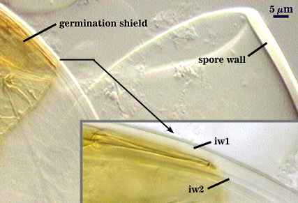
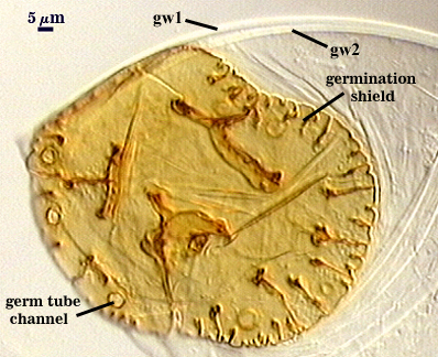
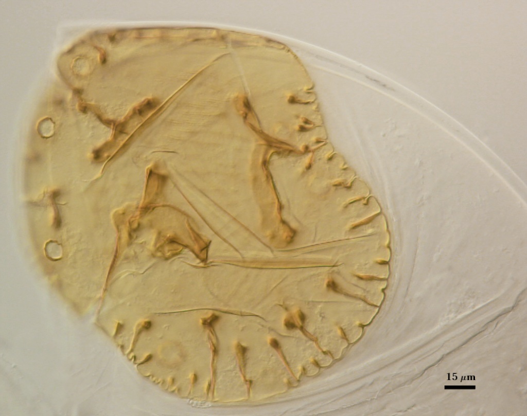
Germination
COLOR: Hyaline to pale yellow-brown (0-10-30-10).
SHAPE: Ovoid, with length approximately 1.5 times that of the width. Margins of shields generally are smooth, with few folds (each with paired germ holes). The shield is sufficiently robust to separate intact from the inner flexible walls when spores are broken. Position of the shield is on iw2.
SIZE: Not measured
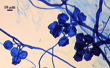
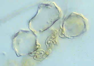
Auxiliary Cells
Aggregate (1-12) cells borne on coiled brown (20-40-80-0) hyphae, thin-walled (< 1 µm thick), pale yellow-brown (0-20-50-10) in transmitted light, each cell with shallow swellings 0.5-2 µm m high and 7-10 µm wide.
| Aggregate Cells | |
|---|---|
 |  |
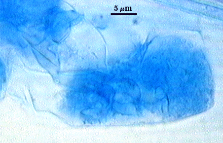
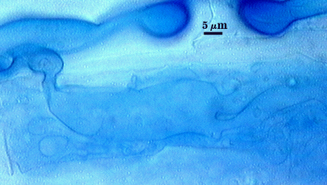
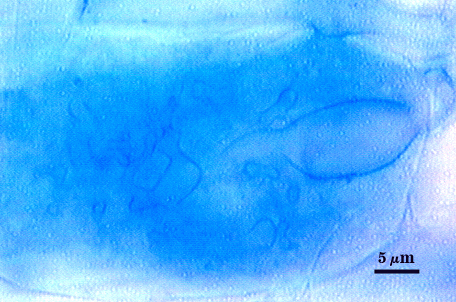
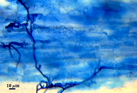
Mycorrhizae
Intraradical arbuscules and hyphae consistently stain darkly in roots treated with trypan blue. Arbuscules with many fine tips from a swollen trunk. Hyphae often with knobs or projections, usually densely coiled near entry points. Auxiliary cells and external hyphae often amass around the root, with the former most abundant in pot cultures just prior to spore formation and declining thereafter.
| Arbuscules in red clover roots | ||
|---|---|---|
 |  |  |
| Mycorrhizae in red clover (with entry points) | |
|---|---|
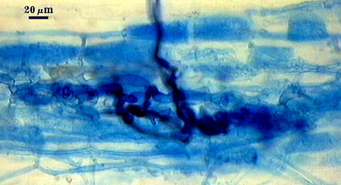 |  |
Notes
Older spores resemble those of S. calospora under a dissecting microscope, except they are more oblong and larger in size range. The original description of S. pellucida (Koske and Walker, 1986) illustrates the problems in interpreting what constituted “walls” and “wall groups” (Walker, 1983).
The images below can be uploaded into your browser by clicking on the thumbnail or can be downloaded to your computer by clicking on the link below each image. Please do not use these images for other than personal use without expressed permission from INVAM.
High Resolution Images | |
|---|---|
 |  |
 |  |
 In Melzer's Reagent | 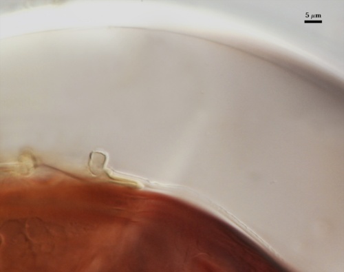 |
Reference
- Koske, R. E. and C. Walker. 1986. Species of Scutellospora (Endogonaceae) with smooth-walled spores from maritime sand dunes: Two new species and a redescription of the spores of Scutellospora pellucida and Scutellospora calospora. Mycotaxon 27:219-235.
- Walker, C. 1983. Taxonomic concepts in the Endogonaceae: spore wall concepts in species descriptions. Mycotaxon 18: 443-455.