Racocetra persica
(reference accession MA461A)
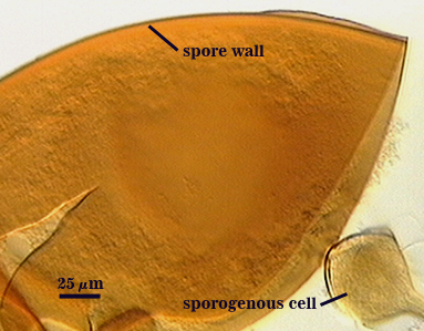
Whole Spores
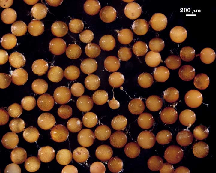
 COLOR: Pale to dark copper (0-20-40-0) to slighly darker cream color (20-60-70-10).
SHAPE: Globose to subglobose.
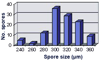
SIZE DISTRIBUTION: 240-360 µm, mean = 313 µm (n = 115).
Subcellular Structure of Spores
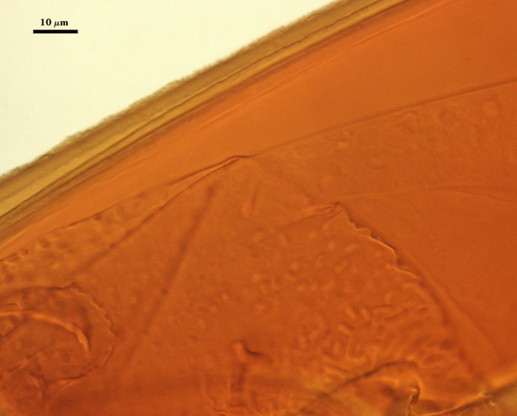
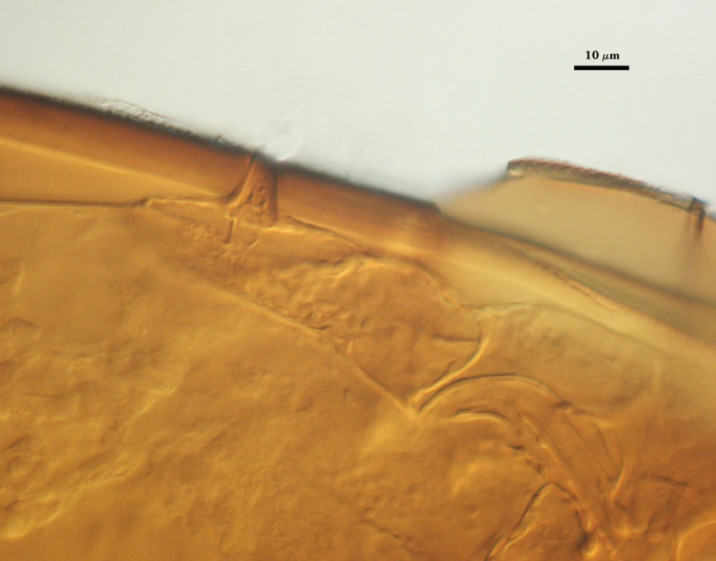
SPORE WALL: Two layers (L1 and L2) that are adherent that in juvenile spores are of equal thickness, with L2 thickening as the spore wall grows and differentiates (sequence in the differentiation of spore subcellular structures—from left to right).
| SPORE WALLS | |||
|---|---|---|---|
 | 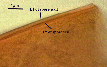 |  | 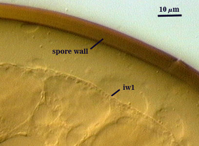 |
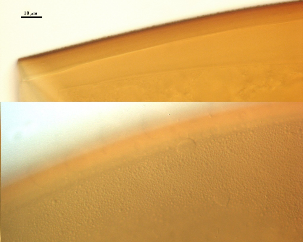
L1: An outer permanent rigid layer, yellow-brown (0-20-80-0), 0.7-1.8 µm thick. The surface consists of many rounded warts 0.5 µm wide and 0.2-0.5 µm high.
| L1 L2 sporewall L2 thicker than L1 | |
|---|---|
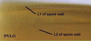 | 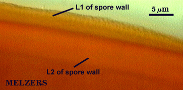 |
L2: A layer consisting of orange-brown (0-30-80-0) to dark orange-brown (0-60-100-0) sublayers (or laminae) that increase in number with thickness, 6.2-8.4 µm thick (mean of 7.6 µm) in mature spores. This layer stains an orange-red (0-60-80-0) to red-brown color (20-80-80-0) in Melzer’s reagent.
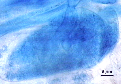
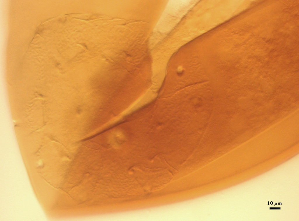
GERMINAL WALLS: One bi-layered hyaline flexible inner wall (gw1) formed independent of the spore wall and subtending hypha.
| GERMINAL WALLS | ||
|---|---|---|
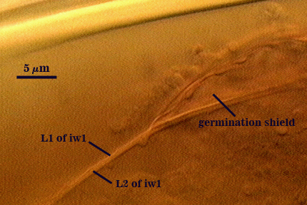 | 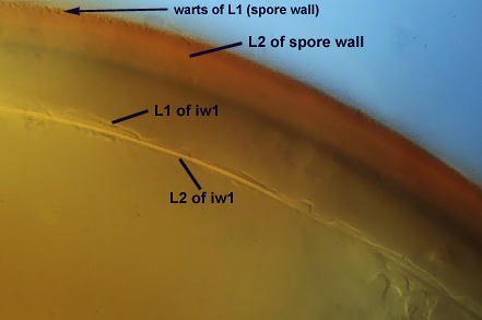 | 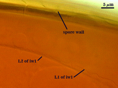 |
GW1: Two layers (L1 and L2) that in field-collected spores are so adherent that they appear as one layer (the condition of type specimens used to describe the species). In spores from pot cultures, the outer layer often separates in small folds from parts of the wall giving the wall a blistered appearance. L1: A thin layer less than 0.5 µm thick and thus difficult to resolve without differential interference optics; no reaction in Melzer’s reagent. L2: A slightly thicker layer, 0.6-1.2 µm thick; no reaction in Melzer’s reagent.
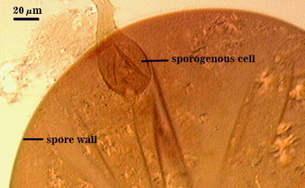
Subtending Hypha
WIDTH OF SPOROGENOUS CELL: 35-46 µm (mean = 42 µm).
SPOROGENOUS CELL WALL: Two hyaline layers (L1 and L2) probably are present (continuous with the two layers of the spore wall), but only L2 is readily discernible at the level of the compound microscope.
L2: Orange-brown (0-30-100-0), 2.4-4.8 µm thick near the spore and then thinning to 1.4-1.6 µm beyond the sporogenous cell.
OCCLUSION: Closure by a plug concolorous with the laminate layer of the spore wall.
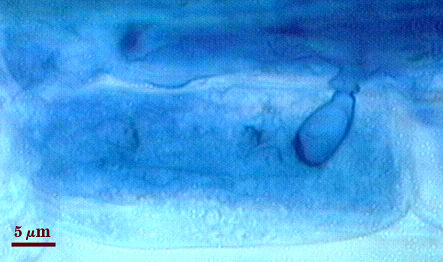
Germination
COLOR: Hyaline to pale yellow (0-0-20-0).
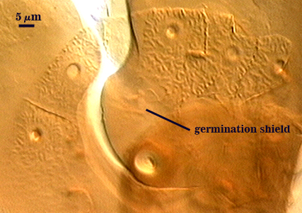
SHAPE: Ovoid, with length approximately 1.5 times that of the width. Shield usually has margins with only shallow convolutions, the surface smooth in some spores (possibly immature?) but with a papillate surface in others that gives it a rugose appearance. The shield forms on the germinal wall.
SIZE: Not measured.
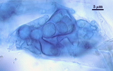
Auxiliary Cells
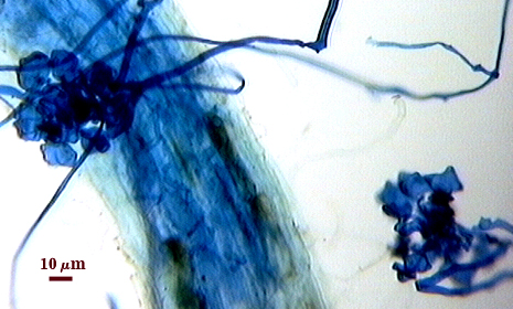
Cells in aggregates of 5-13, subglobose, ovoid to clavate, borne on coiled hyaline hyphae, thin-walled (< 1 µm thick), pale yellow (0-0-10-0) in transmitted light, each cell with tuberculate surface, with swellings 1-5 m high and 3-10 µm wide.
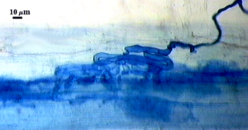
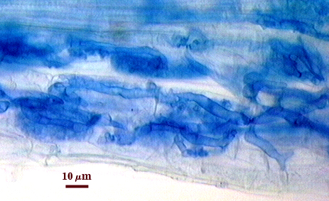
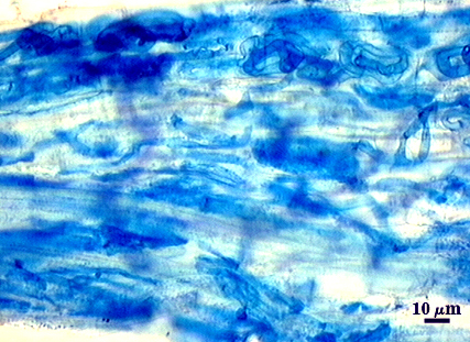
Mycorrhizae
Extraradical hyphae of two morphological types: one wide (3-7 µm) and the other thinner (1.5-2.5 µm). The former usually is the infective hyphae at entry points and forms knobby swellings there and near auxiliary cells. Intraradical arbuscules and hyphae consistently stain darkly in roots treated with trypan blue. Arbuscular hyphae branch to form many fine tips from a swollen basal hypha. Intraradical hyphae 3-11 µm wide, with knobs, projections and swollen areas (up to 14 µm wide), and usually densely coiled near entry points and in outer cortical cells.
| Arbuscule in corn roots | ||
|---|---|---|
 |  |  |
| Mycorrhizal structures in corn roots (with entry point) | ||
|---|---|---|
 |  |  |
Notes
Immature spores are a pale cream (0-0-20-0) to tan with slight rose tint (0-10-40-0 to 0-20-80-0). Ornamentations are fully differentiated at this stage, before many sublayers have formed in the laminate layer. Racocetra castanea is identical to R. persica in all properties except that the outer layer (L1) of the spore wall is smooth.
The images below can be uploaded into your browser by clicking on the thumbnail or can be downloaded to your computer by clicking on the link below each image. Please do not use these images for other than personal use without expressed permission from INVAM.
High Resolution Images | ||
|---|---|---|
|  | |
 |  | |
 | ||