Dentiscutata reticulata
(reference accession CL202)
The culture of this species survived only two propagation cycles before losing all infectivity. The spores were heavily parasitized at the time of deposit (the reason there is no photo of whole spores) and cultures started from individual spores still carried whatever microbe was causing degradation. So this description is from only a few voucher slides that were made and borrows heavily from the protologue.
Whole Spores
COLOR: Dark red-brown (40-80-100-10) to almost black. Immature spores are cream to orange-brown.
SHAPE: Mostly globose.
SIZE DISTRIBUTION: 180-390 µm.
Subcellular Structure of Spores
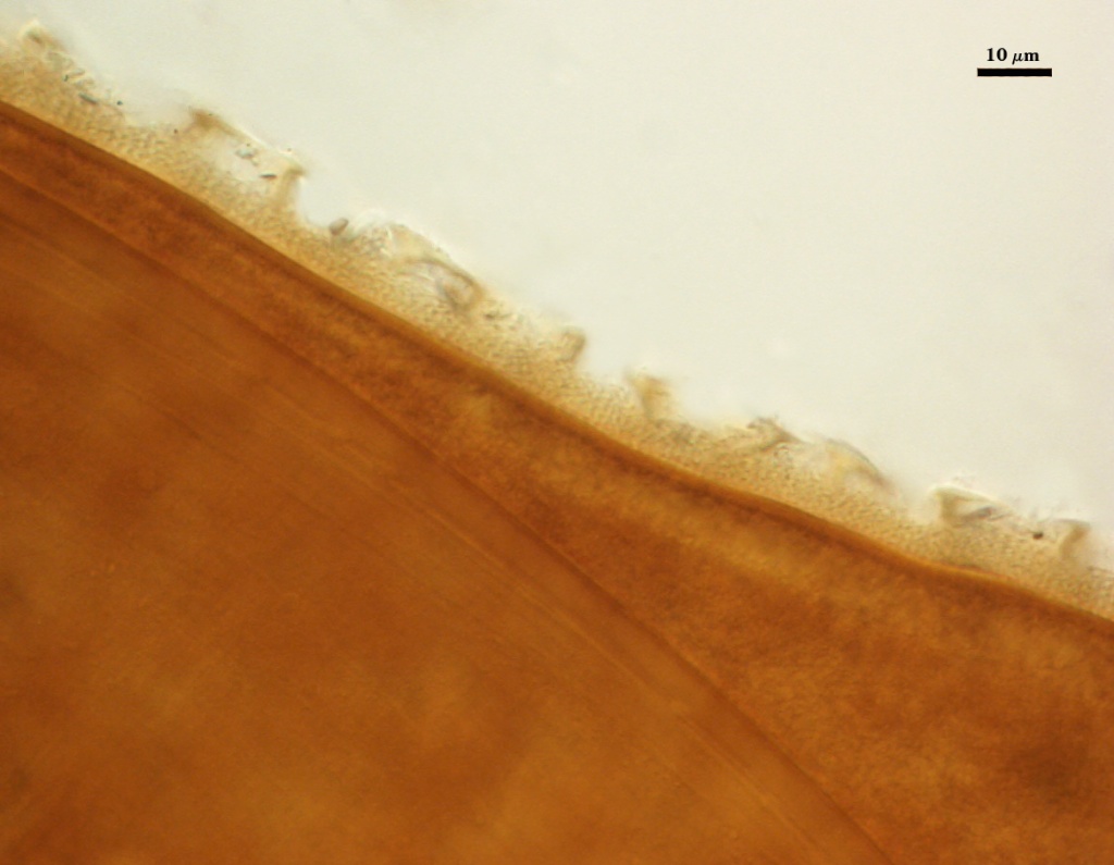
SPORE WALL: Three layers, the outer being ornamented, a middle laminate (thickest) layer, and a very thin inner layer.
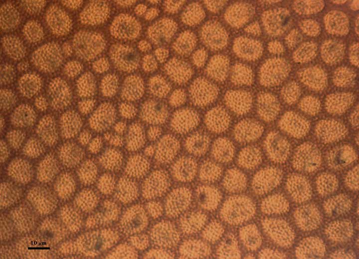
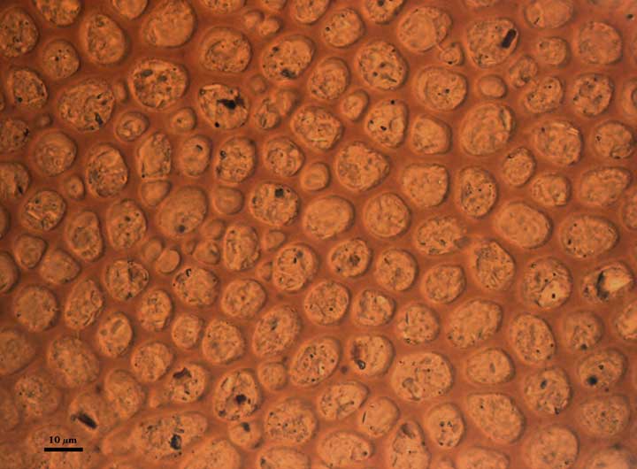
| Reticulated surface | ||
|---|---|---|
|
|
|
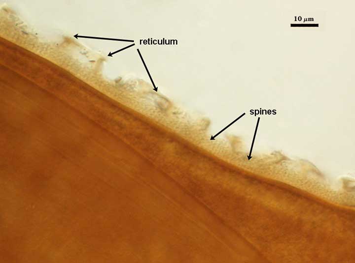
L1: Dark red-brown with a reticulate pattern of polygonal shapes in the center of which are thin densely packed spines.
L2: Laminate layer
L3: Given the similarities with D. nigra, the spore wall also may include a very thin (< 1 µm) inner layer—completely obscured by the outer layers.
GERMINAL WALLS: Two bilayered flexible hyaline inner walls (gw1 and gw2) that are synthesized consecutively after the spore wall has completed differentiation. They are formed after the spore wall as completely differentiated. In many mature spores, both germinal walls tend to stay closely paired, even with applied pressure. They are hard to see in this species because the spore wall is complex and tends to obscure them.
| In PVLG | In Melzer's reagent |
|---|---|
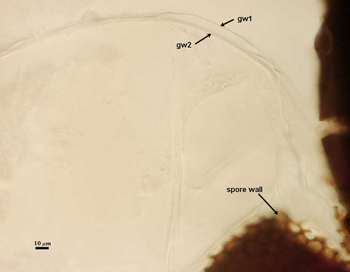 | 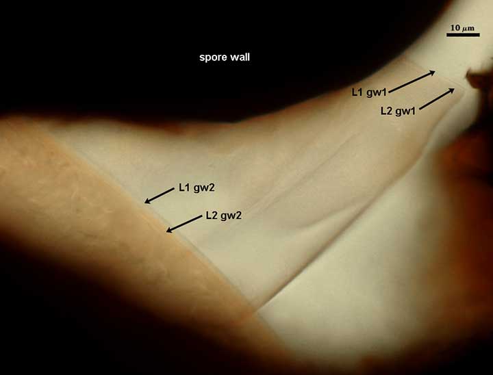 |
GW1: Two layers are formed (L1 and L2) that are tightly adherent. L1 is less than 0.5 µm thick; L2 is slightly thicker (1-2 µm). Both layers are thin enough that when tightly adherent, they appear as one layer.
GW2: Two layers are formed (L1 and L2) that are tightly adherent. The two germinal walls are not easy to separate or distinguish except when a germination shield is formed, which is positioned between the two walls. The inner layer stains light pinkish-red in Melzer’s reagent.
Subtending Hypha
WIDTH OF SPOROGENOUS CELL:
SPOROGENOUS CELL WALL STRUCTURE:
OCCLUSION: Closure by a plug concolorous with L2 of the spore wall.
Germination
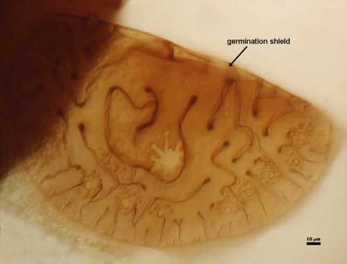
COLOR: Yellow-brown (0-30-70-10).
SHAPE: Circular to ovoid. with an intricate arrangement of invaginations around the perimeter that extend into the center of the structure. Position of the shield is on gw2.
The images below can be uploaded into your browser by clicking on the thumbnail or can be downloaded to your computer by clicking on the link below each image. Please do not use these images for other than personal use without expressed permission from INVAM.
High Resolution Images | |
|---|---|
| |
 |  |
 | |