Gigaspora rosea
(reference accession FL105)
= Gigaspora candida
Whole Spores
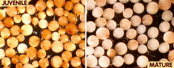
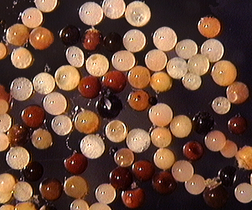
 COLOR: Pale cream with a pale pink tint (0-5-10-0) in new healthy spores. Pale cream (0-0-10-0) to pale yellow-brown (0-5-20-0) in older spores.
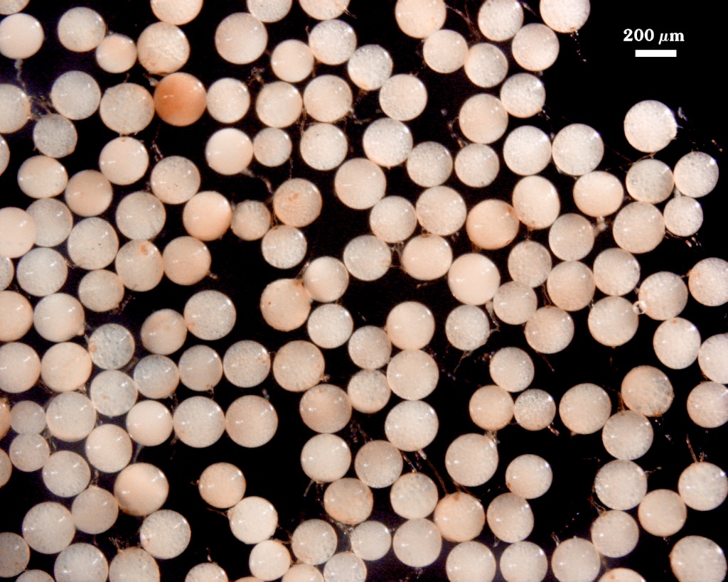
SHAPE: Globose to subglobose.
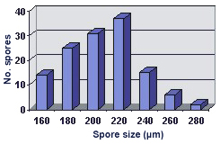
SIZE DISTRIBUTION: 160-280 µm, mean = 206 µm (n = 130).
Subcellular Structure of Spores
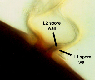
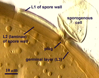
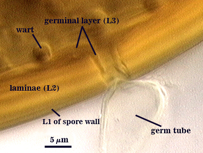
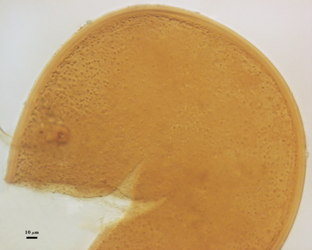
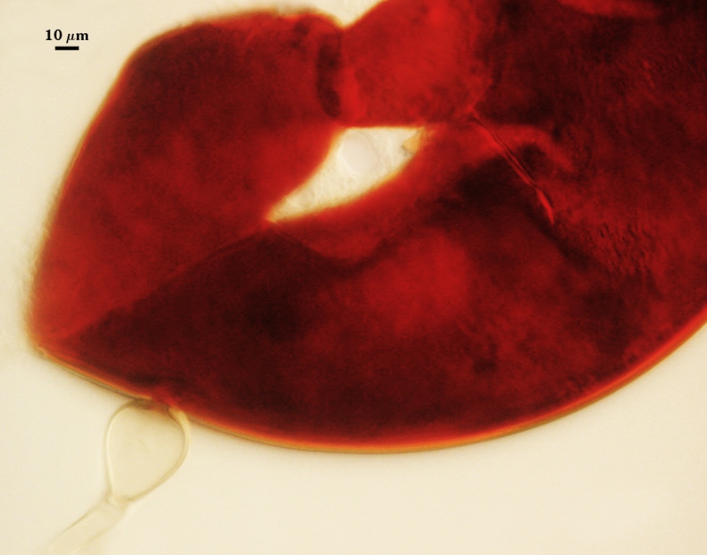
SPORE WALL: Three layers (L1, L2, and L3), the first two adherent and of equal thickness in juvenile spores, with L2 thickening as the spore wall is differentiated and L3 differentiating just prior to germ tube formation.
| In PVLG | In PVLG + Melzer's Reagent |
|---|---|
|
|
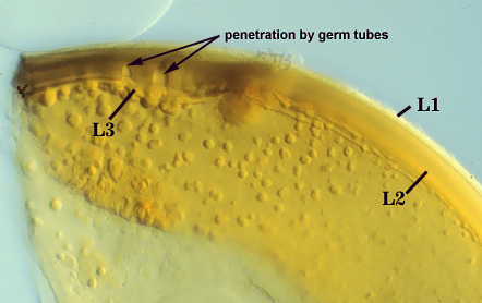
L1: An outer permanent rigid layer, hyaline, 1.5-2.2 µm thick. The surfac3e is smooth, although debris may be attached to older spores, especially those collected from field soils.
L2: A layer consisting of hyaline sublayers (or laminae) that increase in number with thickness; varying considerably in thickness in mature spores; 8-29 µm thick (18-27 µm in just one spore). This variation is attributed to sublayer plasticity that causes spreading as well as breakage with applied pressure to the slide. Sublayers are pale brown (0-10-60-0) in PVLG, staining dark red-brown (20-80-100-0) to a very dark red-purple (60-80-70-10) in Melzer’s reagent.
L3: A “germinal” layer that is concolorous with the laminate (L2) layer. L3 often is most distinct at the ultrastructural level, where it appears electron dense. Under the compound microscope, it is adherent to L2. Numerous “warts” or “papillae” form on the inner surface of this layer, and they are especially concentrated in regions where germ tubes form (usually in close proximity to the suspensor cell); warts 1.2-5 µm high in germinating spores.
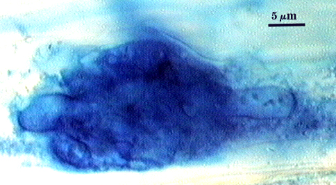
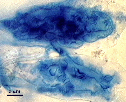
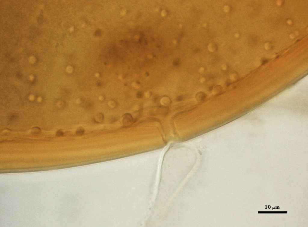
Subtending Hypha
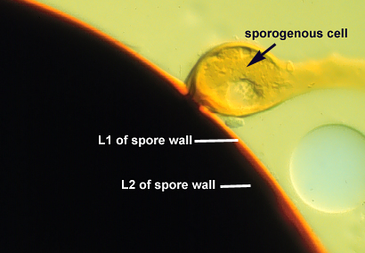
WIDTH OF SPOROGENOUS CELL: 32-45 µm (mean = 39 µm)
SPOROGENOUS CELL WALL: Two hyaline layers (L1 and L2) are evident only in Melzer’s reagent (see photo below), which are continuous with the two layers of the spore wall. In PVLG, but only L2 is easily seen under a compound microscope.
L2: Pale brown (0-10-20-0), 2.0-4.5 µm thick near the spore and then thinning to 1.2-1.4 µm beyond the sporogenous cell.
OCCLUSION: Closure by a plug concolorous with the laminate layer of the spore wall.
| Rosea Spore Wall | |
|---|---|
|
|
Germination
Germ tube forms in vicinity of warty protruberances on inner surface of L3 of the spore wall, hole through spore wall 8-15 µm wide after emergence from the spore wall; the germ tube holes are 8-10 µm wide (see photo below).
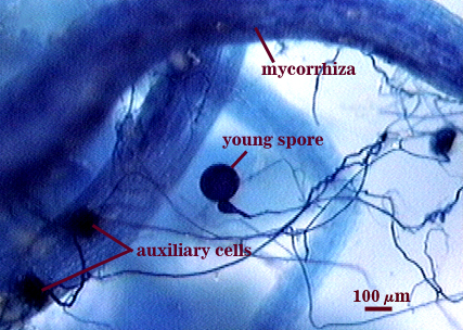
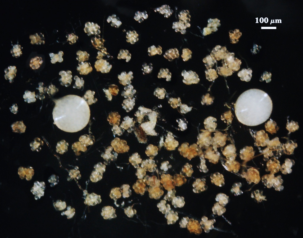
Auxiliary Cells
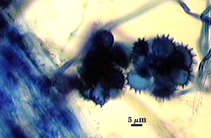
Cells in aggregates of 4-20, subglobose to ovoid to clavate, borne on tightly coiled hyaline hyphae, thin-walled (<1 m thick), hyaline to pale cream (0-0-10-0); each cell with narrow projections 1.5-2.0 µm wide and 2.0-10.0 high.
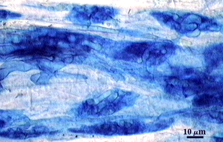
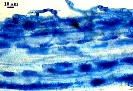
Mycorrhizae
Intraradical arbuscules and hyphae consistently stain darkly in roots treated with trypan blue. Arbuscules produce fine-branches from a swollen basal hypha that is easiest to observe once tips begin to degrade. Intraradical hyphae 3-10 µm in diameter (most between 4-6 µm), with inflated areas up to 16 µm and knob-like projections distributed along length; usually densely coiled near entry points.
| Arbuscules in corn root cortical cells | ||
|---|---|---|
|
|
|
| Arbuscules in corn root cortical cells | |
|---|---|
|
|
Notes
Immature spores are salmon-colored with a slight pink tint (0-10-20-0 to 0-20-60-0). The two layers of the spore wall (L1 and L2) are equally thick (1.6-2 µm). Spores collected from the field can be dark brown to black from parasitism and colonization of the spore lumen by a wide range of soil saprobic microorganisms (see photo at right). Both size and color can separate this species from Gi. margarita if the spores are examined shortly after extraction.
Gigaspora candida was merged with Gi. rosea (Bentivenga and Morton, 1995) when no discrete (discontinuous) morphological differences could be distinguished between the two species.
The images below can be uploaded into your browser by clicking on the thumbnail or can be downloaded to your computer by clicking on the link below each image. Please do not use these images for other than personal use without expressed permission from INVAM.
High Resolution Images | |
|---|---|
 |  |
 |  |
 | |
Reference
- Bentivenga, S. P. and J. B. Morton. 1995. A monograph of the genus Gigaspora incorporating developmental patterns of morphological characters. Mycologia 87: 720-732.