Scutellospora scutata
(reference accession BR243)
Whole Spores
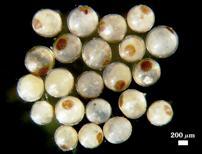
 COLOR: Hyaline/white in most recently formed spores to yellow-brown (0-5-40-0) in older spores (especially those from field soils).
SHAPE: Globose, subglobose, often elliptical or strongly oblong.
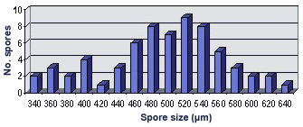
SIZE DISTRIBUTION: 340-640 µm (mean = 494, n = 66)
Subcellular Structure of Spores
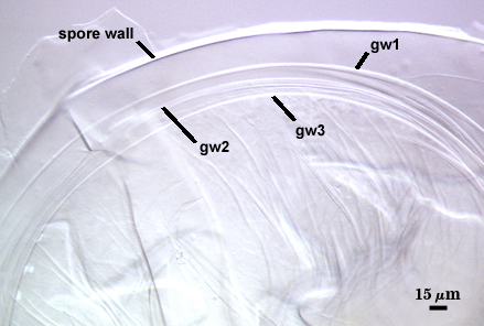
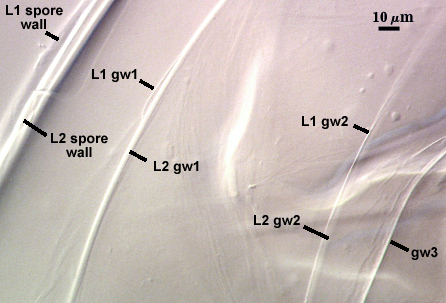
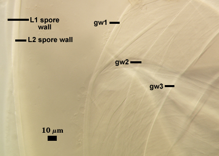
SPORE WALL: Two permanent adherent layers (L1 and L2). Both layers differentiate together.
L1: A single hyaline layer < 1µm thick with a smooth surface.
L2: A rigid layer consisting of fine tightly adherent sublayers (or laminae), 3.5-16 µm thick, white to pale yellow (0-0-10-0) in color.
L3 (?): To see this layer the region of attachment between spore wall and sporogenous cell wall must be intact, and this is extremely difficult to obtain in broken spores. It is assumed to be there because it is present in other related Scutellospora species (most obvious in S. cerradensis).
| Spores mounted in PVLG | |||
|---|---|---|---|
 |  |  |  |
| Spores mounted in PVLG and Melzer’s reagent (1:1 v/v) | |||
|---|---|---|---|
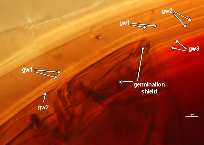 | 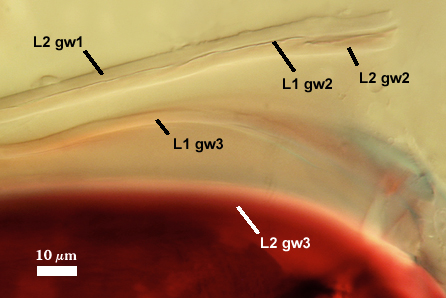 | 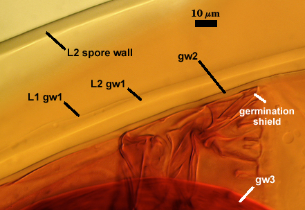 | 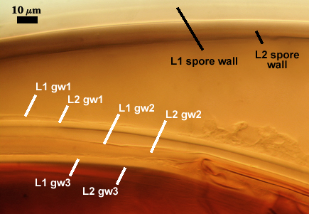 |
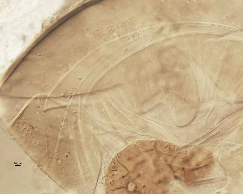
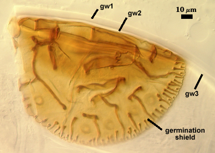
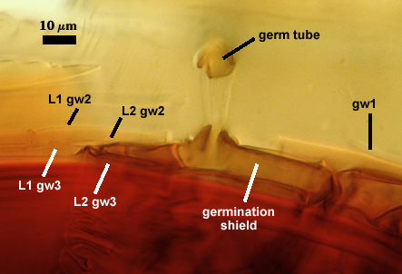
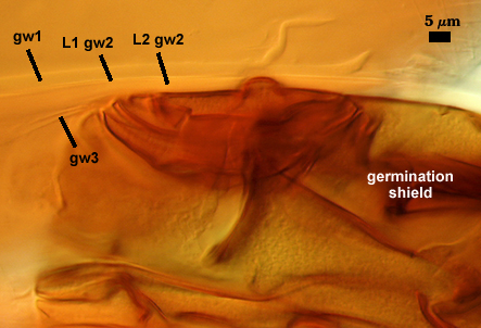
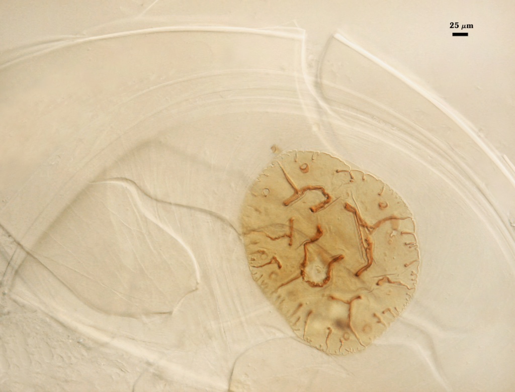
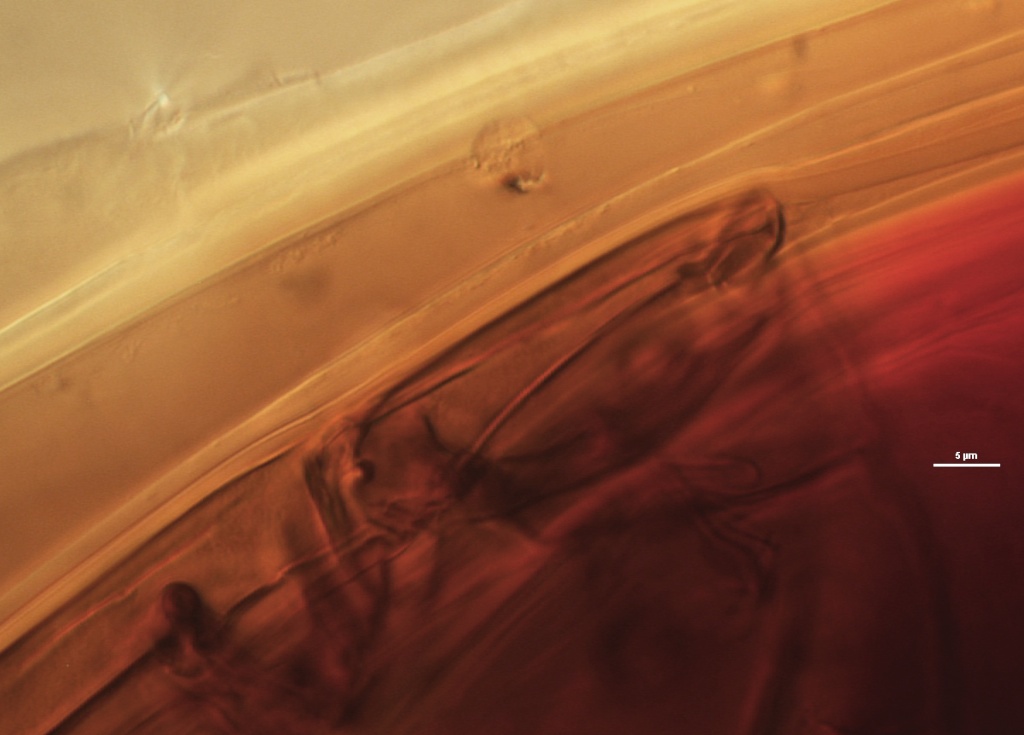
GERMINAL WALLS: Three bi-layered flexible hyaline inner walls (gw1, gw2, and gw3) are present that are completely separate from the spore and sporogenous cell walls. Interpretation of germinal walls can be difficult in broken spores because of difficulties in obtaining a “clean” squash and walls tend to stick together (appearing as one) or fold over each other. Wall number and structure were ascertained most confidently from close examining of inner wall structure at the edges of germination shields (which are always positioned on the innermost germinal wall).
GW1: Two layers (L1 and L2) that usually are adherent; occasionally separable near the broken edge (see photo at right in immature spore in which only gw1 has formed) or with deep folding. L1 is < 0.5 µm thick; L2 is 4-9 µm thick (some of this may be due to swelling with applied pressure). Neither layer reacts in Melzer’s reagent.
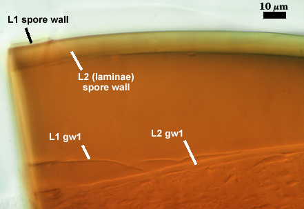
GW2: Two layers (L1 and L2) that also usually are adherent, although they separate more frequently than those of gw1. L1 is < 0.5 µm thick, not reactive in Melzer’s reagent. L2 is slightly plastic, 3-6 µm thick, often producing a pink to light brownish-pink reaction in Melzer’s reagent.
GW3: Two layers (L1 and L2) that usually are adherent, separating when spores are severely crushed. L1 is 3-5 µm thick; slightly reactive in Melzer’s reagent, producing a light pink reaction. L2 is more plastic (or “amorphous”) in newly-mature healthy spores, staining red-purple (20-80-20-0) to dark red-purple (40-80-60-0) in Melzer’s reagent; becoming more rigid with concomitant reduction in staining intensity (to a dark red-brown color) with age and senescence; thickness is highly variable because of this plasticity, ranging from 4.0-18.0 µm thick when amorphous, 3-6 µm thick when more rigid.
Subtending Hypha
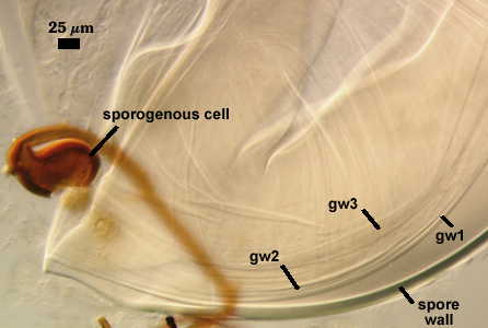
WIDTH OF SPOROGENOUS CELL: 47-90 µm (mean = µm).
SPOROGENOUS CELL WALL: Two layers (L1 and L2) probably are present (continuous with the two layers of the spore wall), but only L2 is discernible under the compound microscope.
L2: Brownish yellow (0-20-80-0), 3-12 µm thick near the spore and then thinning to 1-3 µm beyond the sporogenous cell.
OCCLUSION: Closure by a plug concolorous with the wall of the sporogenous cell.
Germination
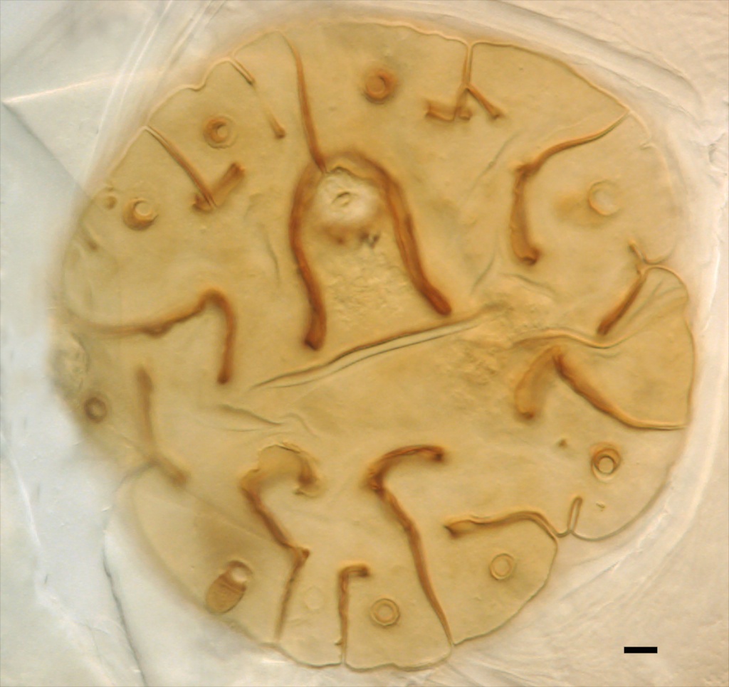
COLOR: Dark yellow-brown (0-30-100-0)
SHAPE: Circular, occassionally more oval-shaped, with multiple lobes; formed on gw3.
SIZE: 240-325×208-302 µm
| GERMINATION SHIELD | |||
|---|---|---|---|
 |  |  |  |
Auxiliary Cells
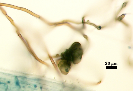
Aggregate (1-12) cells borne on coiled light brown (0-40-80-0) hyphae, thin-walled (< 1 µm thick), pale brown (0-40-80-0) to dark brown (0-60-100-10) in transmitted light, each cell with shallow swellings < 1-2 µm high and 6-10 µm wide.
Mycorrhizae
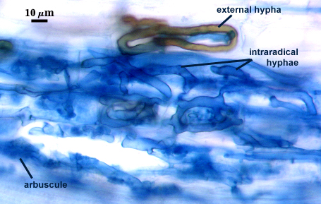
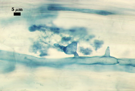
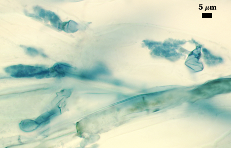
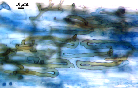
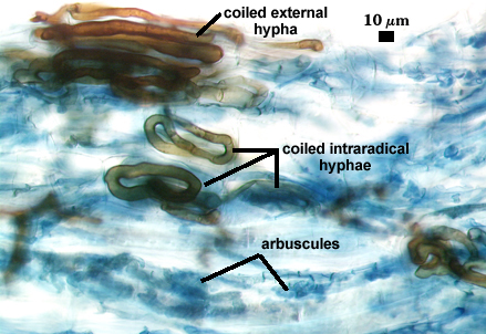
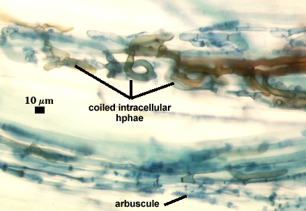
Intraradical arbuscules and hyphae consistently stain darkly in roots treated with trypan blue. Arbuscules with many fine tips from a swollen trunk. Hyphae often with knobs or projections, usually densely coiled near entry points. Auxiliary cells and external hyphae often amass around the root, with the former most abundant in pot cultures just prior to spore formation and declining thereafter. External hyphae are brown.
| Arbuscules in corn roots | ||
|---|---|---|
 |  |  |
| Other mycorrhizal structures in corn | |||
|---|---|---|---|
 |  |  |  |
Notes
Walker and Diederichs (1989) only describe two germinal walls in spores of S. scutata. Given the difficulty in crushing such large spores and the resultant jumble of flexible inner wall structures, such a conclusion is not surprising. We have had this fungus in culture for almost a decade and have mounted numerous spores to find some that gave a clear picture of germinal wall organization.
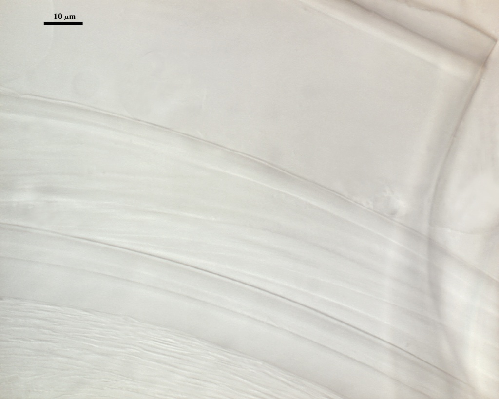
The images below can be uploaded into your browser by clicking on the thumbnail or can be downloaded to your computer by clicking on the link below each image. Please do not use these images for other than personal use without expressed permission from INVAM.
High Resolution Images | |
|---|---|
 |  |
 |  |
 |  |
Reference
- Walker, C. and C. Diederichs. 1989. Scutellospora scutata sp. nov., a newly described endomycorrhizal fungus from Brazil. Mycotaxon 35:357-361.