Acaulospora spinosa
(reference accession WV861A)
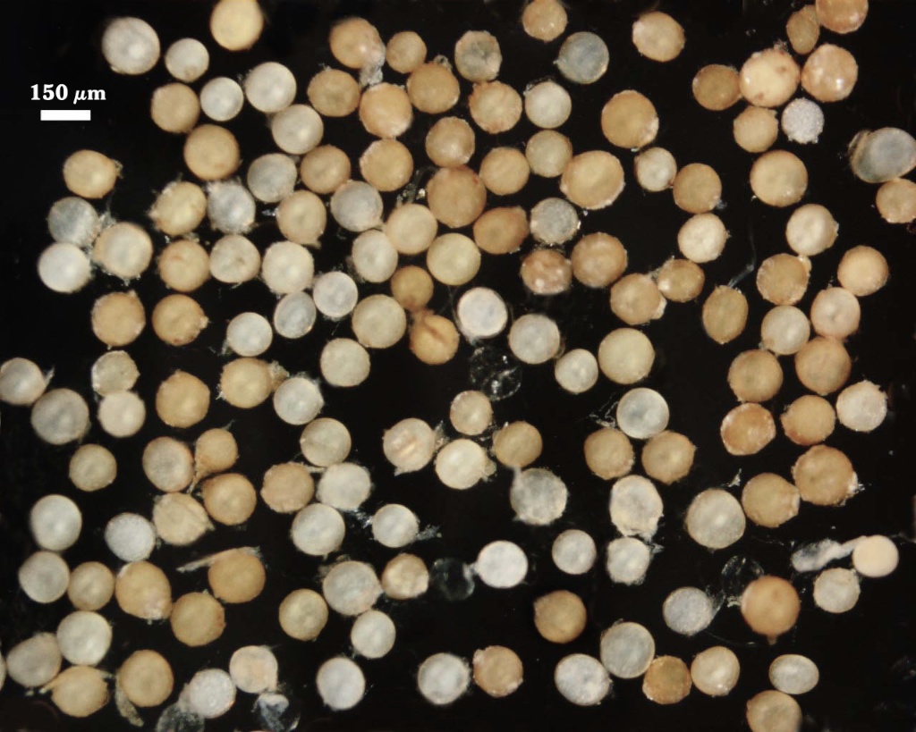
Whole Spores
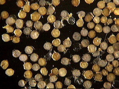
 COLOR: Cream (0-10-20-0) to pale orange-brown (0-30-60-0), with most light yellow-brown (0-10-40-0) in color.
SHAPE: Globose or subglobose.
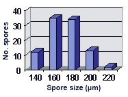
SIZE DISTRIBUTION: 140-220 µm, mean = 171.3 µm (n = 96).
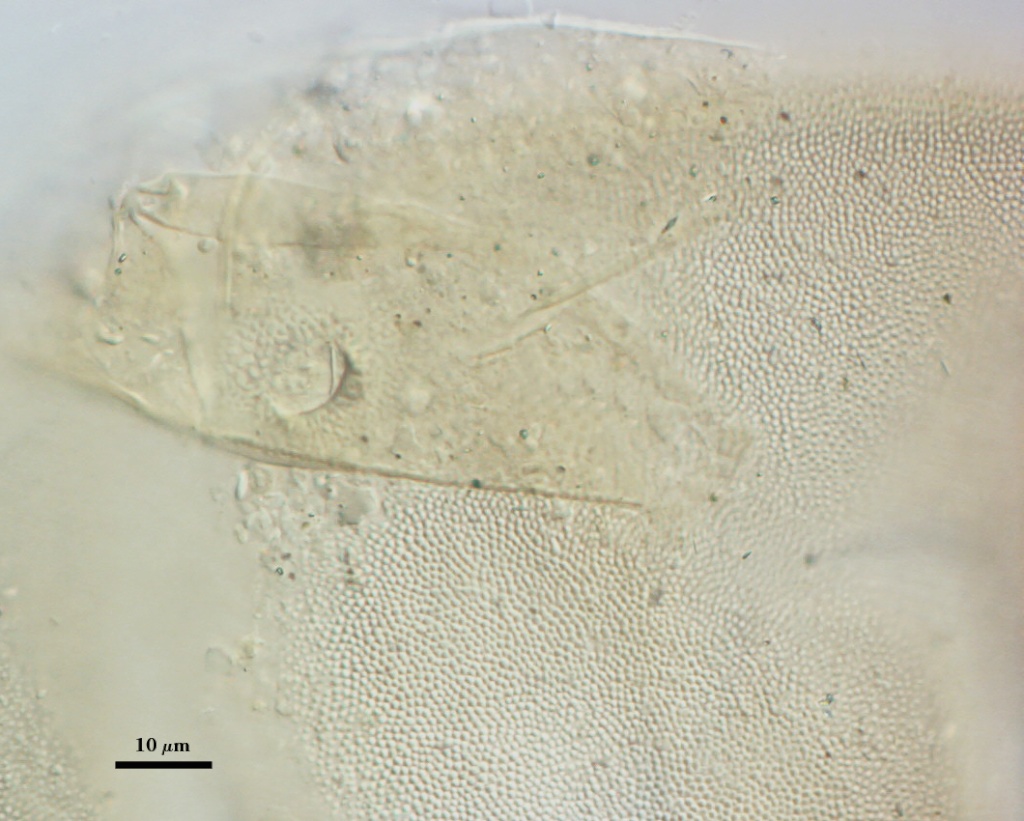
Subcellular Structure of Spores
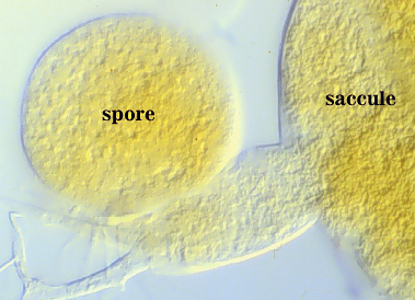
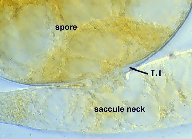
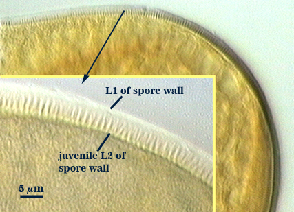
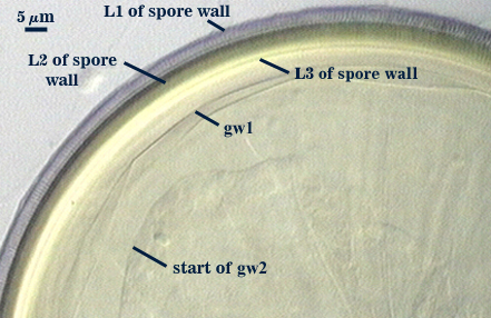
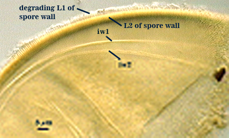
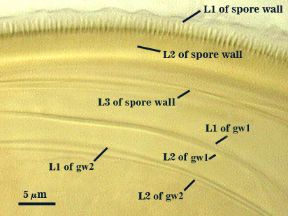
SPORE WALL: Two layers (L1 and L2), the outer continuous with the wall of the neck of the parent sporiferous saccule and the latter being synthesized with origin of the spore (see in sequence of differentiation of spore wall layers and of the two flexible inner germinal walls below, from left to right).
| Differentiation of spore wall layers | |||
|---|---|---|---|
 |  |  |  |
 |  |  | |
L1: Hyaline, 1.2-1.6 µm thick. It degrades or sloughs very early in spore wall differentiation and is absent even before spines on L2 (below) are fully formed.
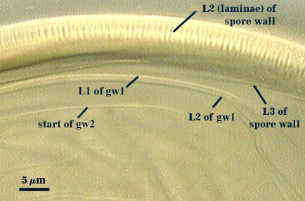
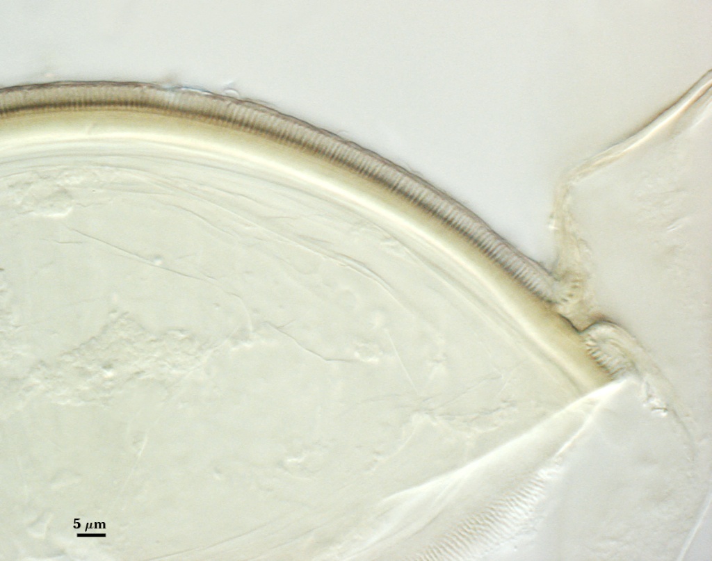
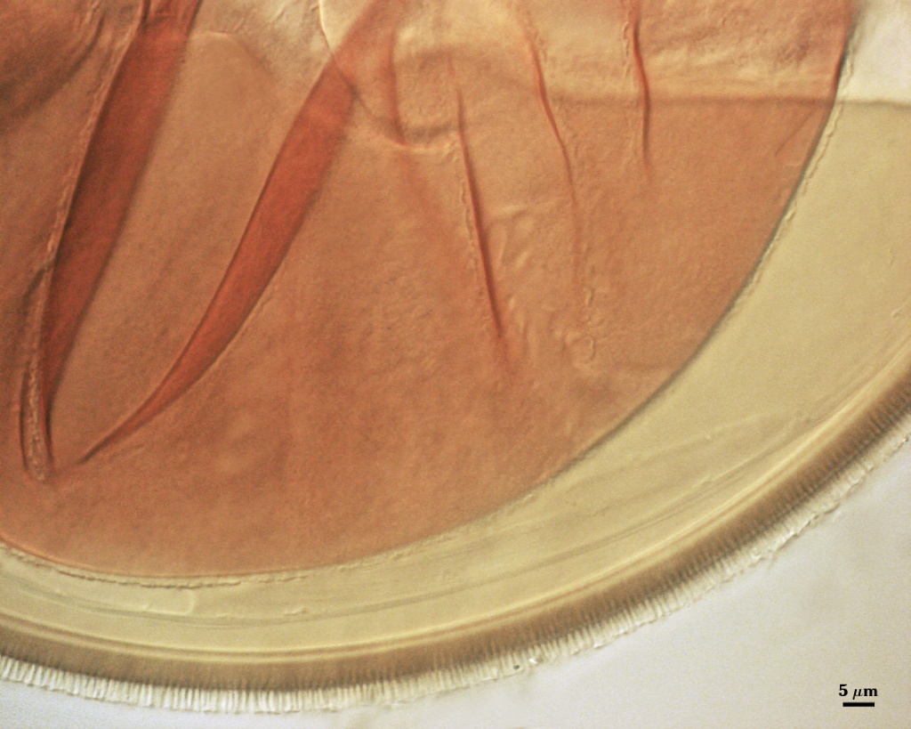
L2: A layer that thickens initially by formation of pale yellow (5-0-30-0) sublayers (or laminae) followed by synthesis of closely packed (< 0.5 m apart) rounded spines 0.5-0.7 m wide and 2-3 m long. Composite thickness of this layer (sublayers + spines) is 6-8.5 m (mean of 7.2 µm).
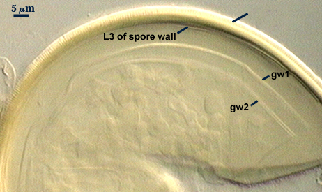
L3: A single hyaline layer, 0.6-1.2 µm thick either is adherent to L2 (and thus hard to see) or more often slightly separable (where it resembles a flexible inner wall). L3 tends to be detected more often in mountants containing Melzer’s reagent because there is a greater degree of separation from the spore wall.
| Spores mounted in PVLG | |
|---|---|
|
|
| Spores in PVLG & Melzer’s reagent (1:1 v/v) | |
|---|---|
|
|
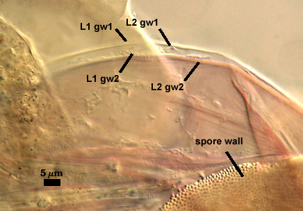
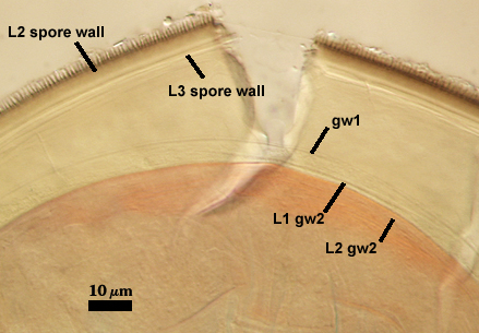
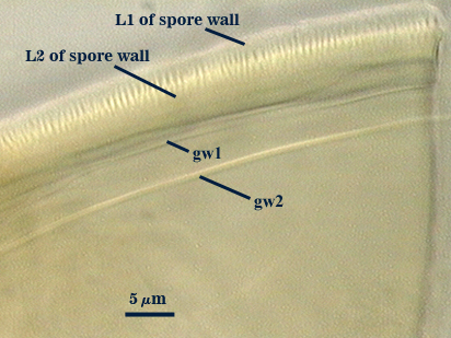
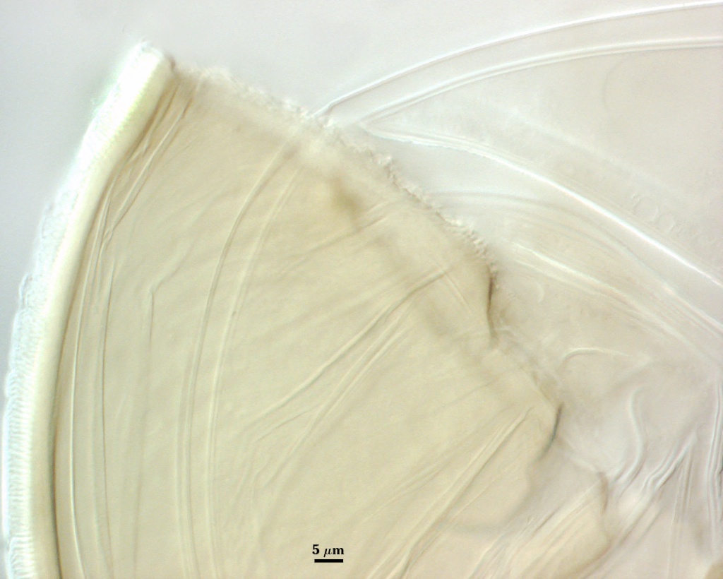
GERMINAL WALLS: Two flexible hyaline inner walls (gw1 and gw2) can be seen in all spores IF they separate when each spore is broken. Layers of the first germinal wall at one time were described as separate “semi-rigid unit walls” (Walker, 1983) because they often broke with the spore wall whereas the more flexible gw2 remained intact (see photo below).
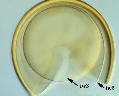
GW1: Two layers (L1 and L2) with a composite thickness of 0.9-1.2 µm. L1 is less than 0.5 µm thick. L2 may be of equal thickness or slightly thicher. This wall readily separates from the spore wall, and thus is consistently seen in broken spores.
GW2: A hyaline wall consisting of two layers (L1 and L2). L1 is 0.6-1.2 µm thick and covered with granular excrescences (“beads”) that tend to dissociate in broken spores or disappear after several months of storage. L2 is 1-1.4 µm thick and stains pink (0-30-20-0) to purplish-pink (0-20-60-40-0) in Melzer’s reagent.
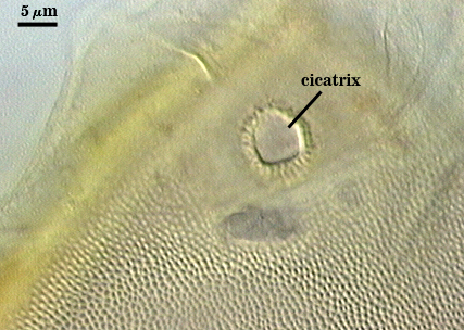
Cicatrix
Scar indicating region of contact between spore and saccule neck during spore synthesis; diameter is 8-19 µm (mean = 11.8 µm ). The edges of the scar are prominent because the attachment between spore and saccule neck consists of a short branch (or pedicel).
Sporiferous Saccule
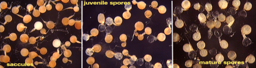
COLOR: Hyaline at maturity. Usually pink to salmon colored in juvenile stages (see sequence of photos below).
SHAPE: Mostly globose.
SIZE DISTRIBUTION: 100-140 µm, mean =129 µm
SACCULE WALL: Two layers, 1.8-3.2 µm thick together, both of near equal thickness (1.1-1.6 µm); sometimes appearing as a single layer.
DISTANCE FROM SACCULE TO SPORE: 50-90 µm (most 80 µm).
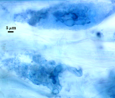
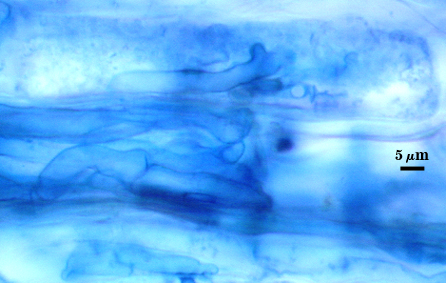
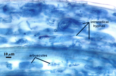
Mycorrhizae
Arbuscules and intraradical hyphae stain faintly in trypan blue, although intensity can be highly variable with age of the mycorrhizae and host plant. Infection units appear to be patchily distributed with oblong to irregular vesicles often forming in small clusters.
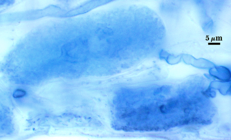
| Arbuscule in cortical cells of a corn root | ||
|---|---|---|
 |  |  |
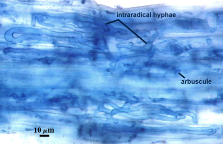
| All mycorrhizal structures in corn roots | ||
|---|---|---|
 |  |  |
Notes
Spore is borne on a short pedicel (3-4 µm long) from the neck of the saccule. The subtending hyphal width at the cicatrix is much broader than that of the cicatrix (28-45 µm). Proportion varies frrom 0.2-0.6 µm (cicatrix-hypha), with a mean of 0.38 µm.
High Resolution Images | |
|---|---|
 |  |
 |  |
 | 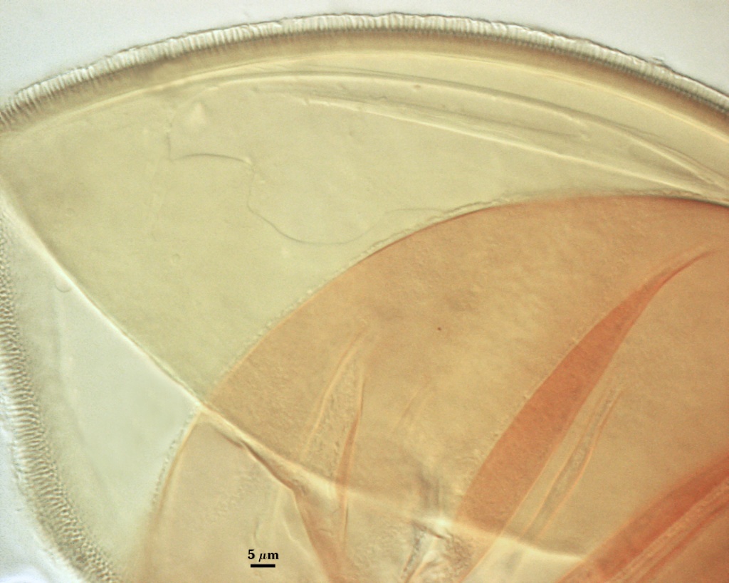 |