Acaulospora sporocarpia
Whole Spore
This description is a combination of information obtained from the protologue (Rothwell and Trappe, 1979), type specimens, and universal patterns of morphological organization and structure in Acaulosporaceae. This species was described from herbarium specimens and has yet to be characterized from healthy living tissue.
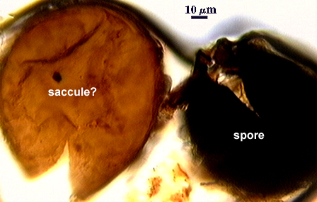
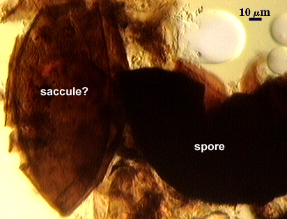
Sporocarps irregular, dark-brown, up to 2.5×1.5×1.5 cm, composed of spores that appear black in incident light, plus sporogenous saccules, hyphal swellings, hyphae and debris.
Spores formed laterally on hypha subtending a single sporiferous saccule. Spores ellipsoid, broadly fusiform, subglobose,or globose, (140-) 160-200 (-240) x (125-) 150-175 (-200) µm diam, dark brown or black, borne on a single pedicel branching from the neck of the saccule.
Subcellular Structure of Spores
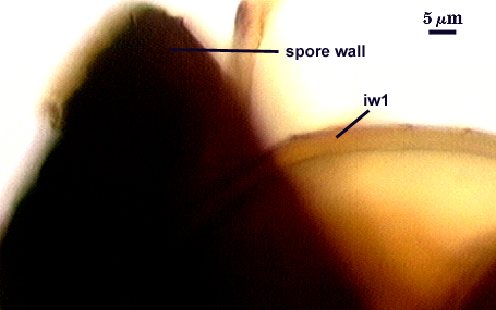
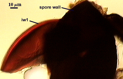
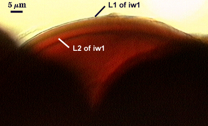
The spore wall is composed of two separable groups: the outer consists of a single wall (wall 1), dark reddish brown in transmitted light, 2.5-5 um thick, laminate, with a rough outer surface; the inner consists of a single wall (wall 2), hyaline, 7-15 µm thick, tough, laminate, and sometimes separating into 2 or more layers when crushed.
| Spore subcellular structure (in PVLG) | ||
|---|---|---|
 |  |  |
| Spore subcellular structure (in Melzer’s reagent) | ||
|---|---|---|
 |  |  |
Pedicel
The pedicel is continuous with hypha that subtends the sporiferous saccule, may be persistent, brown, up to 40 µm long and 20-30 µm wide at the point of attachment to the spore.
Reference
- Berch, S. M. 1985. Acaulospora sporocarpia, a new, sporocarpic species, and emendation of the genus Acaulospora (Endogonaceae, Zygomycotina). Mycotaxon 23:409-418.