Diversispora tortuosa
(reference accession JA301B)
=Glomus tortuosum
Whole Spores
| Sporocarp | |
|---|---|
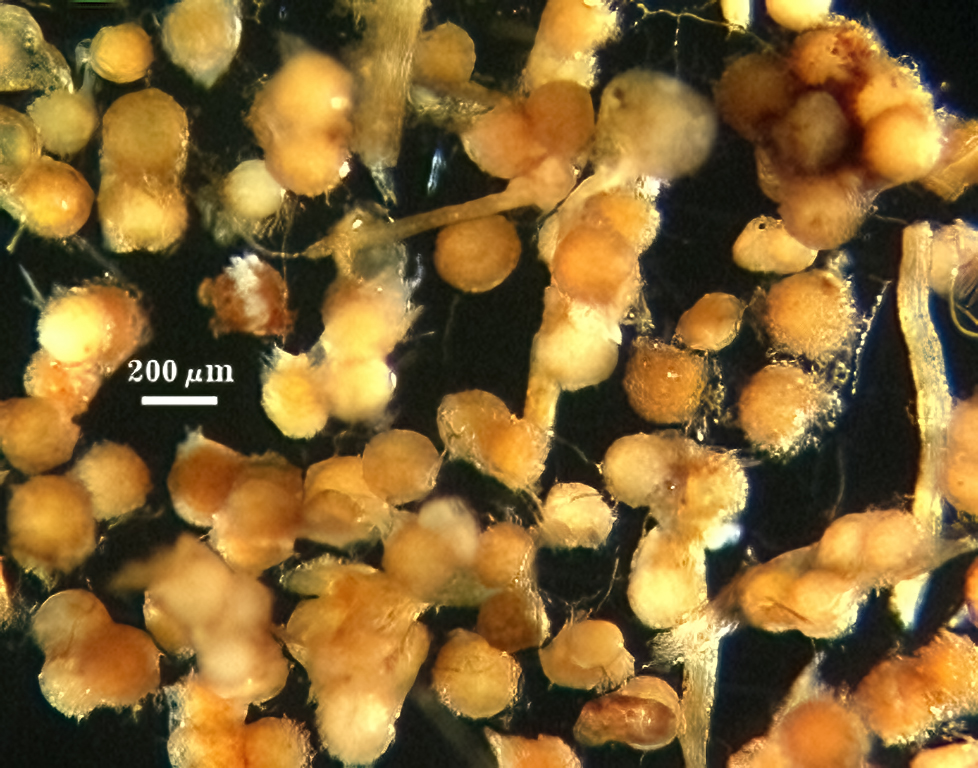 |  |
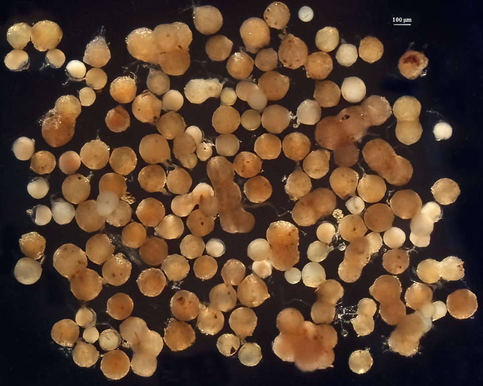
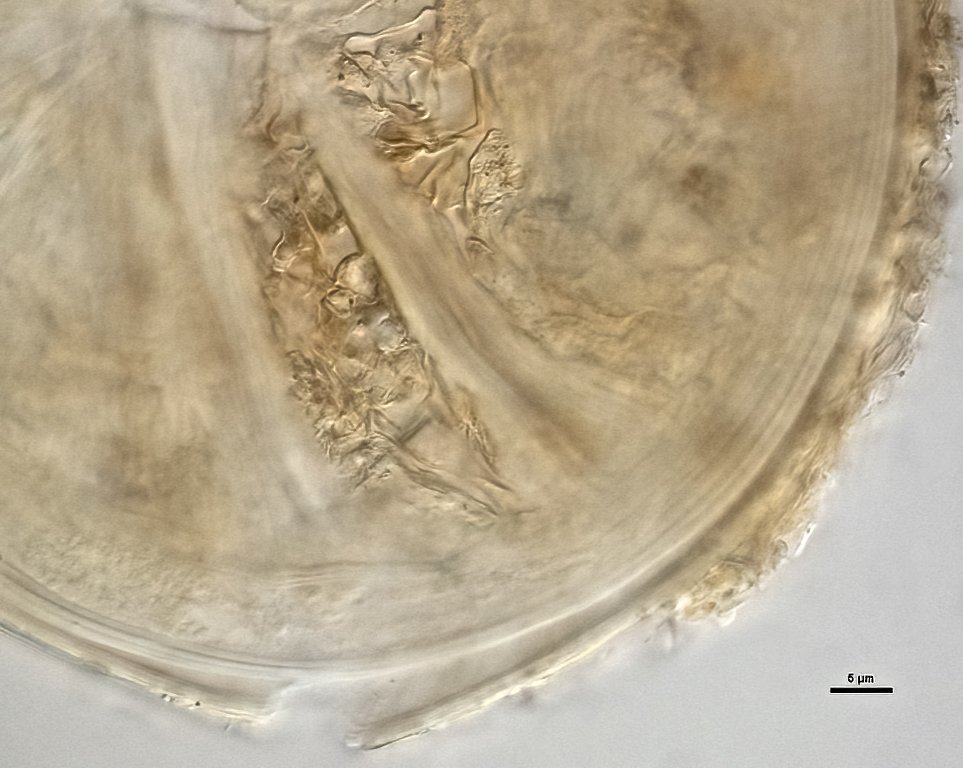
 COLOR: Spores are light yellow-brown (0-10-60-0) to orange-brown (0-40-100-10), tortsize.gif (2158 bytes) but most are pale orange-brown (0-20-40-0)
SHAPE: Mostly globose to subglobose; occasionally irregular.
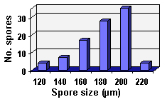
SIZE DISTRIBUTION: 120-220 µm; mean = 181 µm (n = 102)
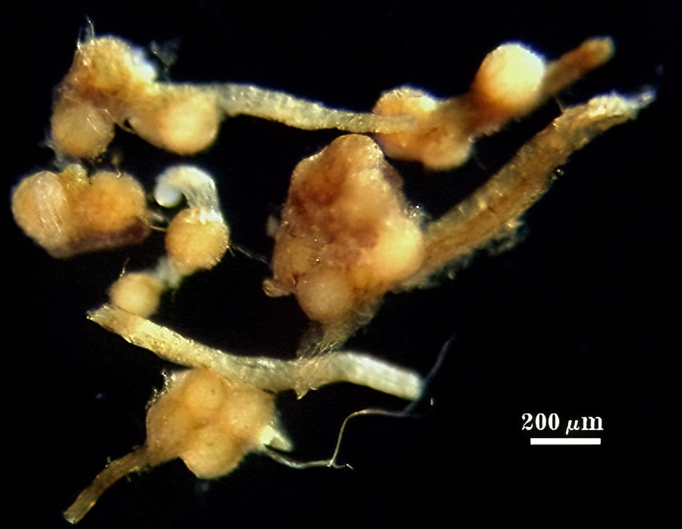
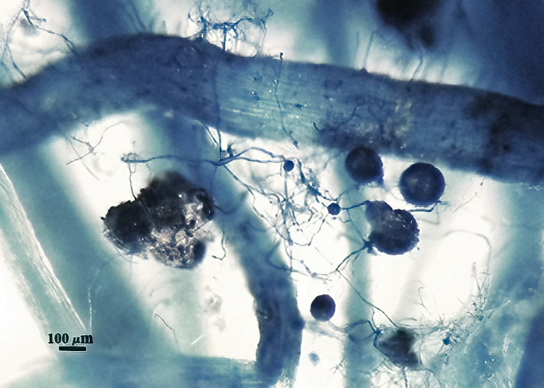
SPOROCARPS: While some spores are formed singly with a peridium covering the spore wall (see below), many spores appear to develop randomly within a dense hyphal peridium. Each sporocarp contains between 2-6 spores and ranges from 290-540 µm in diameter.
Subcellular Structure of Spores
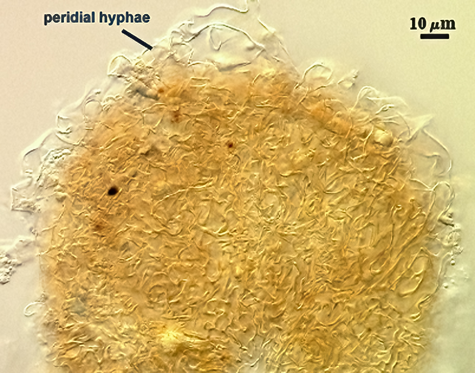
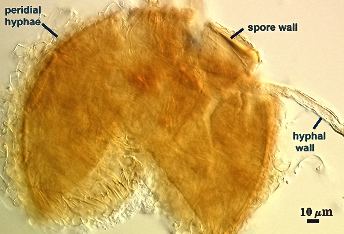
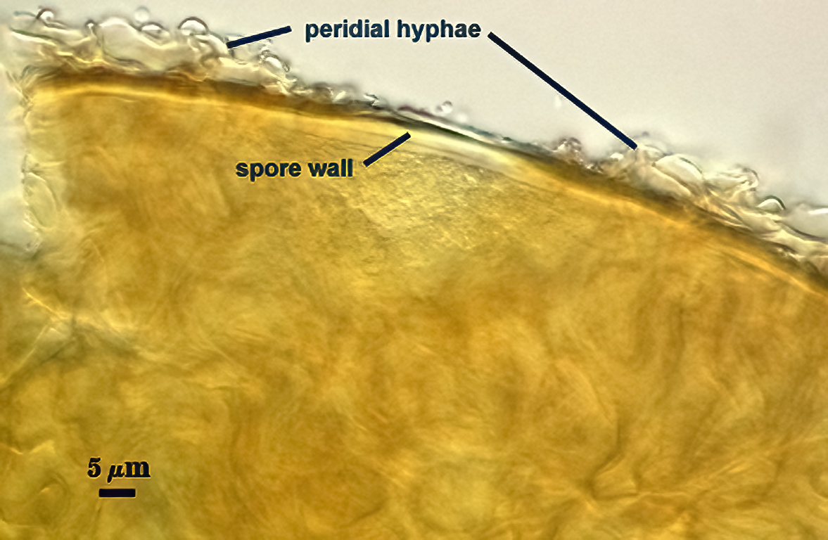
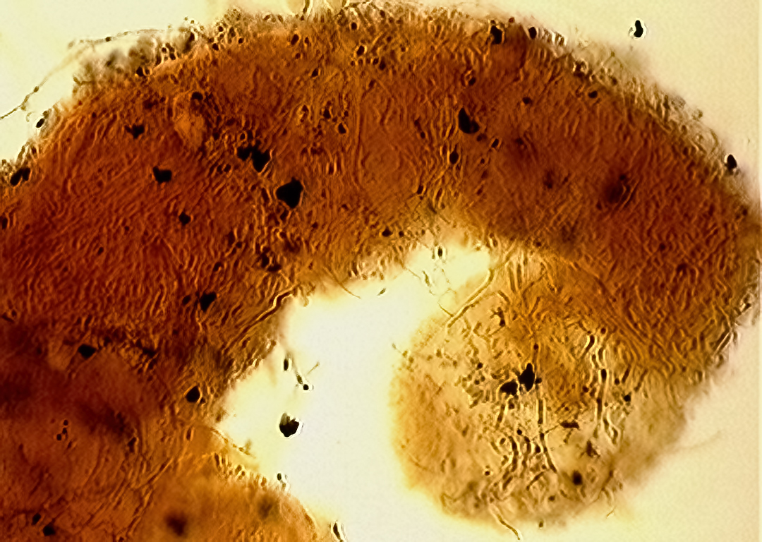
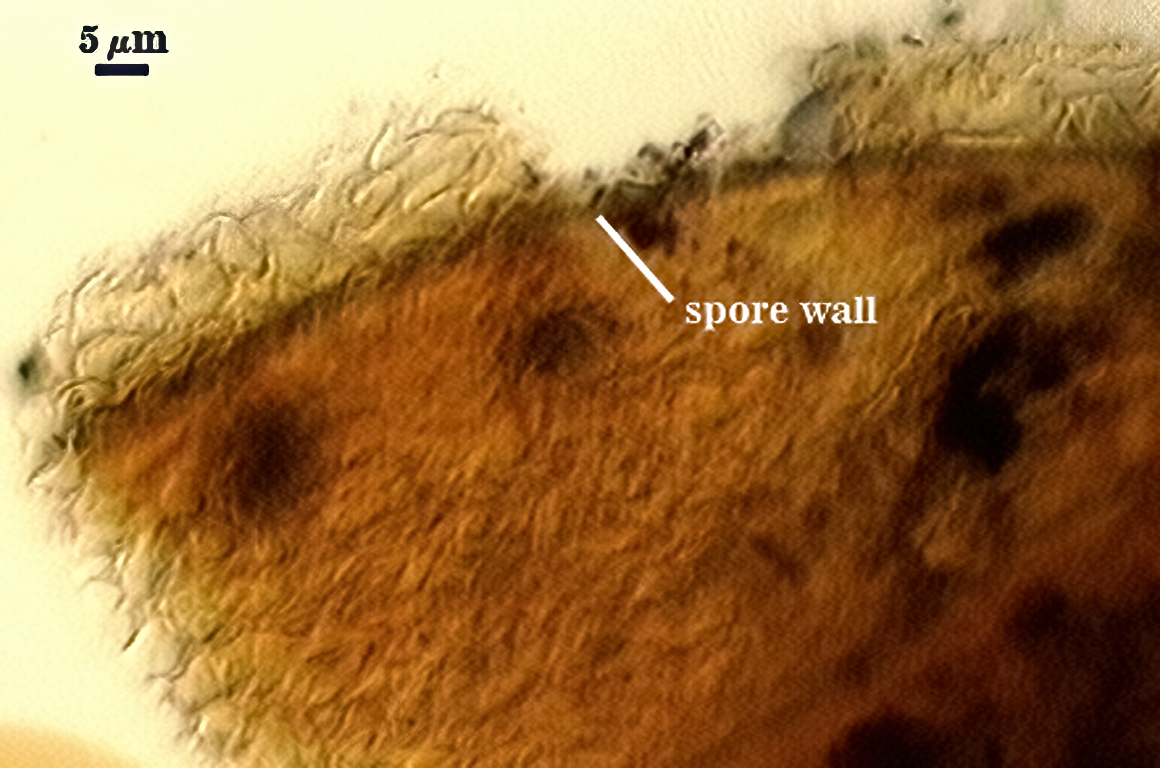
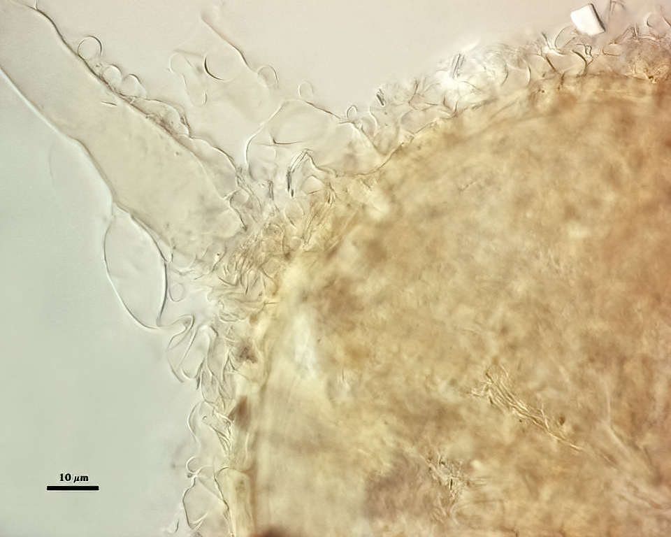
SPORE WALL: One layer (L1) surrounded by dense peridial hyphae.
L1: Pale yellow-brown (0-0-80-0 to 5-0-80-0) sublayers (or laminae) that usually remain adherent, although they may separate at various positions to split the layer into two or three separate components of varying thickness. Thickness varies from µm (mean = µm) in mature spores. This layer originates from the wall of the subtending hypha.
Peridium: Consisting of thin-walled hyphae (walls less than 0.5 µm usually) that are tightly interwoven; they appear to originate from branching of the subtending hyphal wall. The peridium varies considerably in thickness on the spore wall, but rarely exceeds 25 µm.
| Spores mounted in PVLG | ||
|---|---|---|
|
|
|

| In Melzer’s reagent | |
|---|---|
|
|
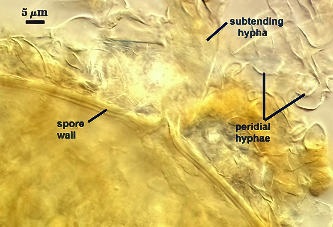
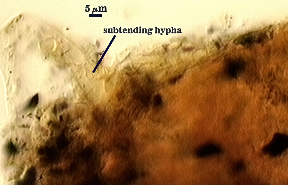
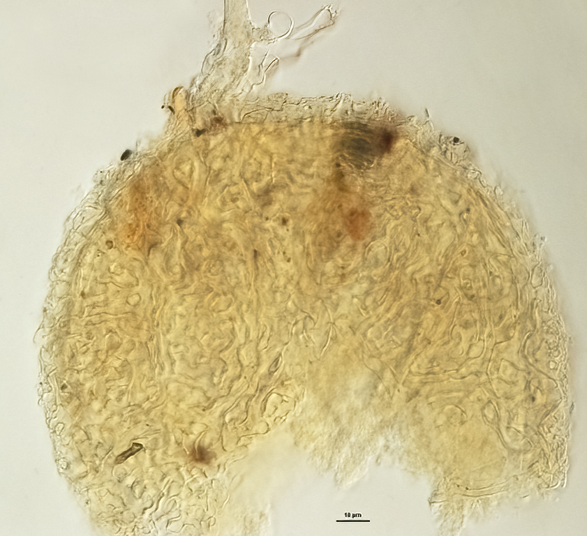
Subtending Hypha
SHAPE: Cylindrical to slightly flared, although shape at the spore often is hard to detect because of interference from surrounding peridial hyphae (see photos above).
WIDTH: 12.5-15 µm (mean = 13.2 µm).
COMPOSITE WALL THICKNESS: 1.5 µm.
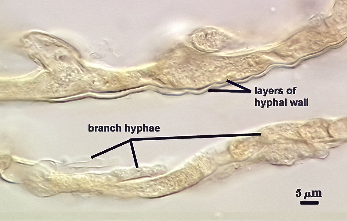
WALL STRUCTURE: Appears to vary from 1-2 layers, but this variation may be confusion on which hyphae near the point of attachment represents the parent hypha and those that branch (with considerably complexity) from that hypha. There appear to be two types of hyphae formed in culture: thicker hyphae (10-15 µm) with two wall layers (outer layer 1 µm thick or less, inner layer 0.5 µm or less) from which branches numerous thinner hyphae (3.8-6 µm) with a single wall layer less than 1 µm thick.It is uncertain which of these two types form sporogenous hyphae, but the former is hypothesized to be the main contender.
OCCLUSION: Not discernible in the spores mounted thus far.
Germination
Not observed.
Notes
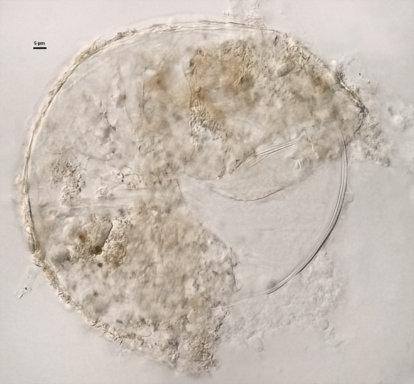
Spores in this culture closely resemble those of type specimens (see photos below), except that they are lighter in color (which is to be expected in fresh versus preserved material).
| Tortuosa cluster | ||
|---|---|---|
 |  |  |
The images below can be uploaded into your browser by clicking on the thumbnail or can be downloaded to your computer by clicking on the link below each image. Please do not use these images for other than personal use without expressed permission from INVAM.
High Resolution Images | |
|---|---|
 |  |
 |  |
 |  |