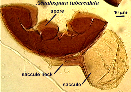
Acaulospora tuberculata
Whole Spores
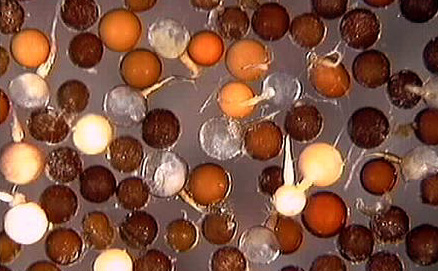
 COLOR: Red-orange (0-50-60-5) to dark red- brown (40-50-70-10).
SHAPE: Globose or subglobose.
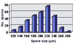
SIZE DISTRIBUTION: 120-280 µm, mean = 202 µm (n = 304).
Subcellular Structure of Spores
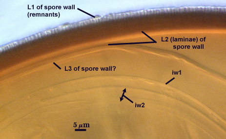
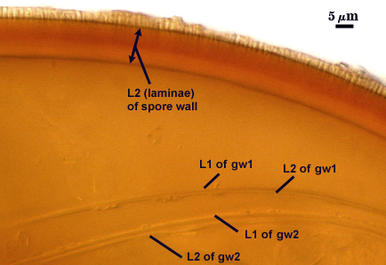
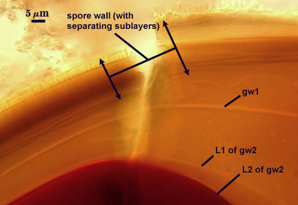
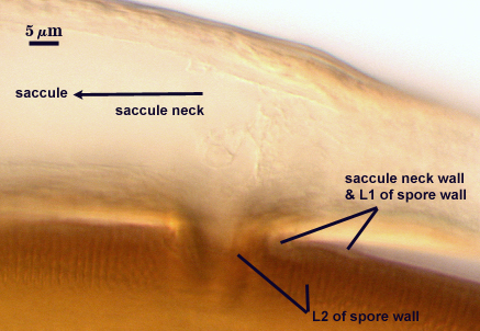
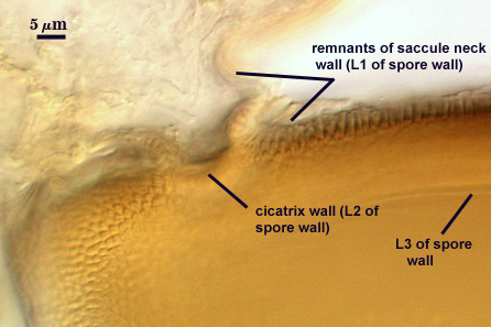
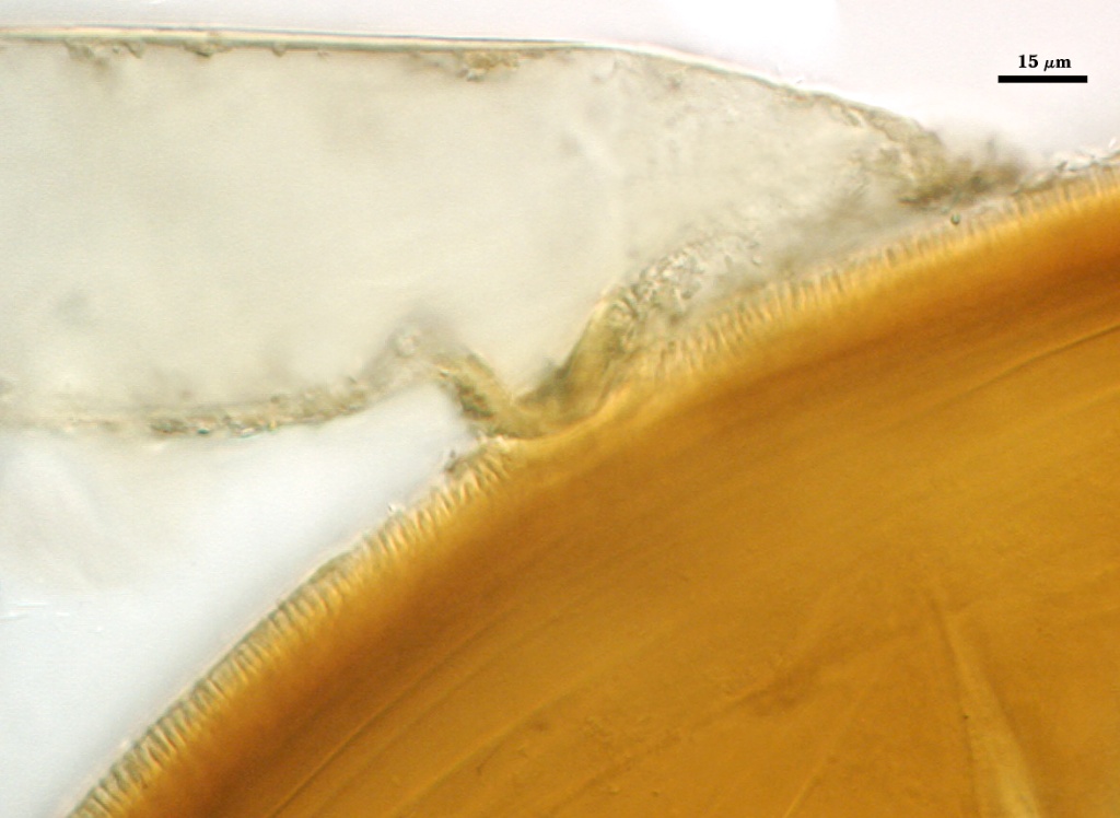
SPORE WALL: Three layers (L1, L2 and L3), the outer layer continuous with the wall of the neck of the parent sporiferous saccule and the latter two are synthesized with development of the spore.
L1: Hyaline, 0.9-3.75 µm thick; remains after tubercles on L2 (below) have formed in some spores, but it is not observed except from a side view of L2.
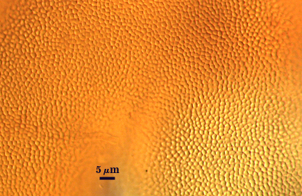
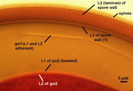
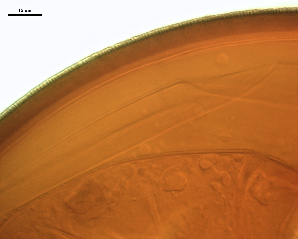
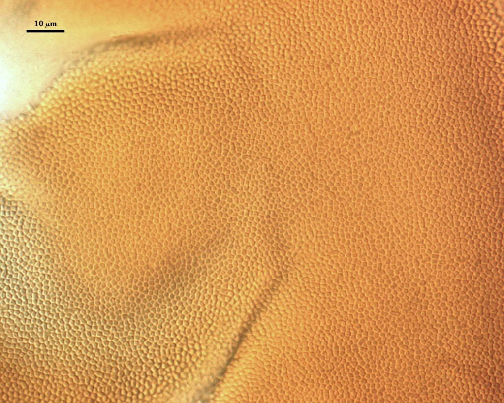
L2: A layer that thickens initially by formation of red-brown sublayers (or laminae) followed by synthesis of polygonal (4-5 sides) spines, or tubercles, 1.4-3.5 µm high and 1.0-1.3 µm wide. Total thickness of this layer (sublayers + tubercles) ranges from 8.5 to11.8 µm (mean = 9.24 µm ). At maturity, the pore between spore and saccule neck is bridged by sublayers of L2 (minus tubercules) to form an “endospore”.
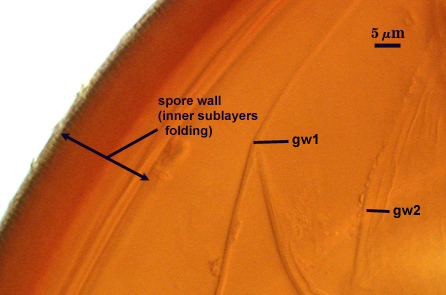
L3: A yellow-brown to red-brown layer which appears discrete in some spores due to separation from the spore wall, but which otherwise appears to be inner sublayers of L2. It can be undetected in some spores (completely adherent to L2), appear as a pair of thin adherent flexible layers, or appear as a wide range of layers due to folds; ranging from 0.5-4.0 µm when measurable.
| Stored spores (> 90 days) mounted in PVLG | ||
|---|---|---|
|
|
|
| Stored spores in PVLG and Melzer’s reagent | |||
|---|---|---|---|
|
|
|
|
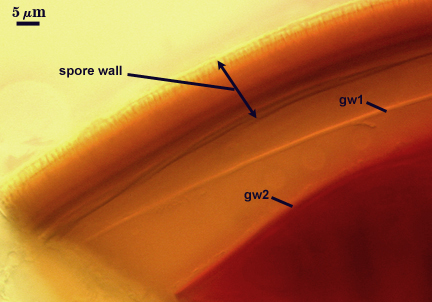
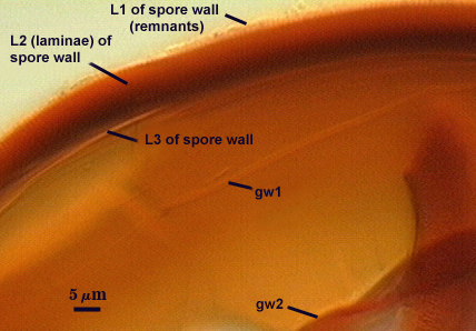
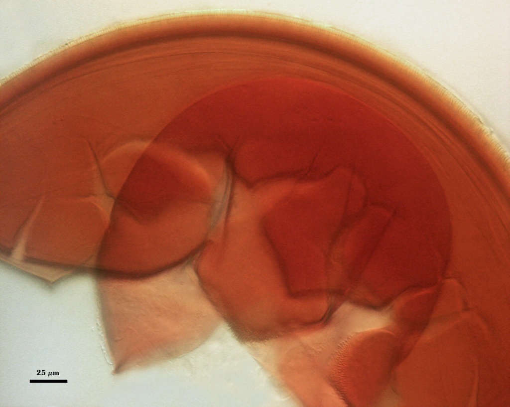
GERMINAL WALLS: Two flexible hyaline inner walls (gw1 and gw2) are present. They can be seen only after spores have completely cleared when mounted in PVLG or Melzer’s reagent, which can take as long as 30 days.
GW1: Consisting of two adherent hyaline layers (L1 and L2) are formed, both are of near-equal thickness, ranging from less than 0.5 µm to 0.8 µm thick. This wall forms completely independent of the spore wall and thus has no linkages with it.
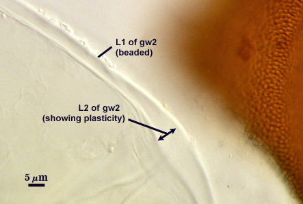
GW2: Consisting of two adherent hyaline layers (L1 and L2) are formed that together are 5-10.6 µm when measured in PVLG. L1 is hyaline, 0.8-1.8 µm thick with granular excresences (or “beads”) that tend to become dislodged and float away when pressure is applied to it. These “beads” are stabilized after preservation in formalin, but otherwise may be absent on mounted spores within a few months of storage. L2 is hyaline and somewhat plastic (probably equivalent to a “coriaceous” wall using traditional terminology). It is 1.2-2.0 µm thick in PVLG-based mountants, staining pinkish red (20/80/60/0) to red-brown (40/80/60/0) in Melzer’s reagent.
Cicatrix
A circular to ovoid scar indicating region of contact between spore and saccule neck; it consists of closely packed tubercles surrounding an unornamented depression, 9-16 µm in diameter (mean = 12.7 µm) at its widest.
Sporiferous Saccule
COLOR: Hyaline.
SHAPE: Mostly globose.
SIZE DISTRIBUTION: 100-140 µm, mean =129 µm
SACCULE WALL: Two adherent layers (L1 and L2), the outer layer often difficult to see. L1 is hyaline, smooth, 1.0-2.4 µm thick on the saccule, increasing to 2.5 µm thick in region of attachment to saccule neck; continuous with the layer that initially is the juvenile spore wall (L1). L2 consists of pale yellow-brown (0-10-20-0) to orange-brown (0-30-60-0) sublayers tightly adherent on the saccule and loosening near subtending hypha; 1.8-3.4 µm thick except in region of attachment to saccule neck, where it increases to 6.4-12.4 µm; continuous with L2 of the spore wall.
SACCULE NECK: Two adherent layers (L1 and L2). L1 is hyaline, smooth, < 0.6 µm thick except in region of attachment to the saccule. L2 consists of orange-brown (0-60-100-10) sublayers that together are 4.2-8.6 µm thick between saccule and spore, then thinning to 1.0-2.4 um distal to the point of attachment.
PEDICEL: This structure connects the neck of the sporiferous saccule with the spore wall and varies in length from 1.6-8.0 µm. It is cylindrical and 11.2-17.8 µm wide. The wall of the pedicel consists of the two layers (L1 and L2) that are continuous between the spore wall and the saccule neck wall.
| Saccule neck wall | |
|---|---|
|
|
DISTANCE FROM SACCULE TO SPORE: 54-96 µm.
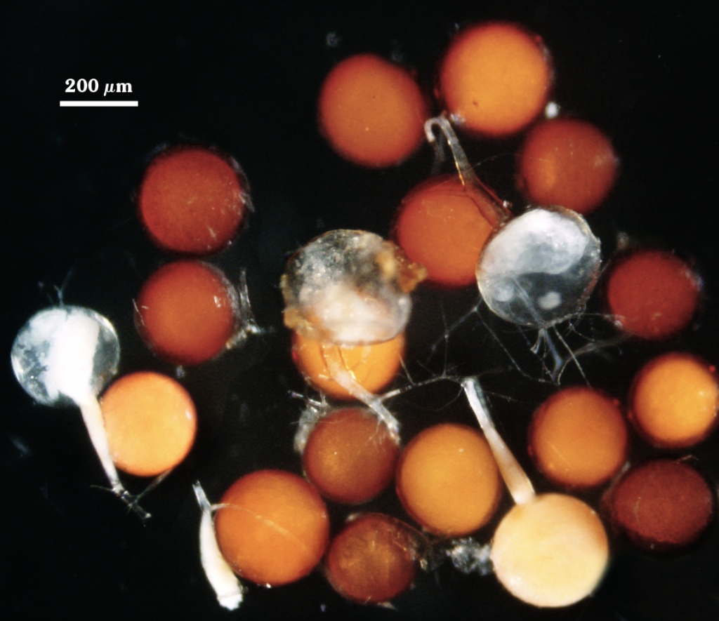
Germination
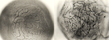
An ovoid “germination orb” forms on iw3 like that found in other Acaulospora species, based on a report by Spain (1992). The photos below were provided courtesy of Joyce Spain.
Mycorrhizae
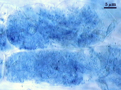
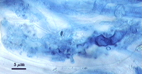
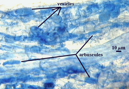
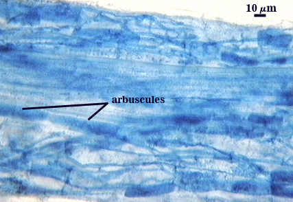
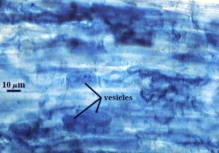
Intraradical arbuscules, vesicles, and hyphae tend to stain with variable intensity in trypan blue, from almost invisible to darkly. Hyphae are coiled mostly near entry points, 3-6 µm in diameter; otherwise thinner (2-4 µm) and usually growing parallel to each other and interconnecting via branches that form “H” and “h” connections. Vesicles often are localized in clusters that are patchily distributed along a mycorrhiza. Many appear to form near entry points (primary or secondary ingress) and many are lobed or irregularly-shaped.
| Arbuscules in corn root cortical cells | |
|---|---|
 |  |
| All mycorrhizal structures in corn root | ||
|---|---|---|
 |  |  |
Notes
Tubercles on the spore wall complete differentiation before any flexible inner walls are formed (or L2 and L3 of the spore wall are fully expanded with sublayer thickenings). The spore forms somewhat like a Glomus species in that the outer layer of the saccule neck (L1) branches and expands for form the initial spore wall, followed by synthesis of a laminate layer in both the parent hypha (saccule neck) and the spore.
The description of this species (Janos and Trappe, 1982) was based on field-collected spores that were largely parasitized, so that few if any germinal inner walls were present or intact. It is not surprising that spores were thought to have only “an easily separable hyaline inner layer” rather than discrete flexible germinal walls as we know them today.
This species proved to be extremely difficult to sporulate in culture. Steve Bentivenga found only a few spores in the original accession (VZ103), inoculated them onto sorghum, took the cone-tainer contents and transplanted to a pot, and then reseeded that pot at least twice in succession to obtain enough colonization for sporulation to occur. The entire process took over 18 months before sporulation was moderately abundant.
High Resolution Images | |
|---|---|
 |  |
 |  |
 | |
Links to Gene Sequences in Genbank
Reference
- Janos, D. P. and J. M. Trappe. 1982. Two new Acaulospora species from tropical Americana. Mycotaxon 15:515-522.