Racocetra verrucosa
(reference accession VA105B)
Whole Spores | |
|---|---|
|
|

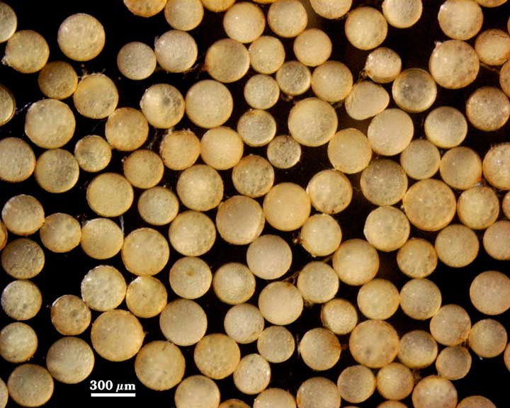
 COLOR: Pale straw (0-10-10-0) to orange brown (0-20-40-0), the former predominant in a healthy culture, the latter more common in old cultures and field collections.
In the right photo above: there is a 50-50 mix of mature and young spores. The latter are easily identified by dense contents and uneven coloration with dark and light regions. The tiny spore at the top is obvious immature as it just begins to expand in size.
SHAPE: Globose, subglobose.
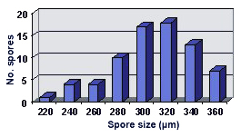
SIZE DISTRIBUTION: 220-360 µm, mean = 308 µm (n = 74).
Subcellular Structure of Spores
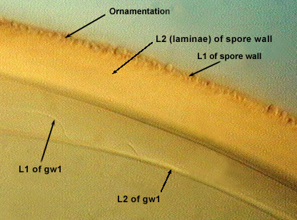
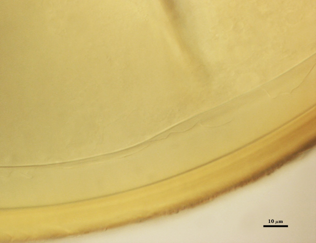
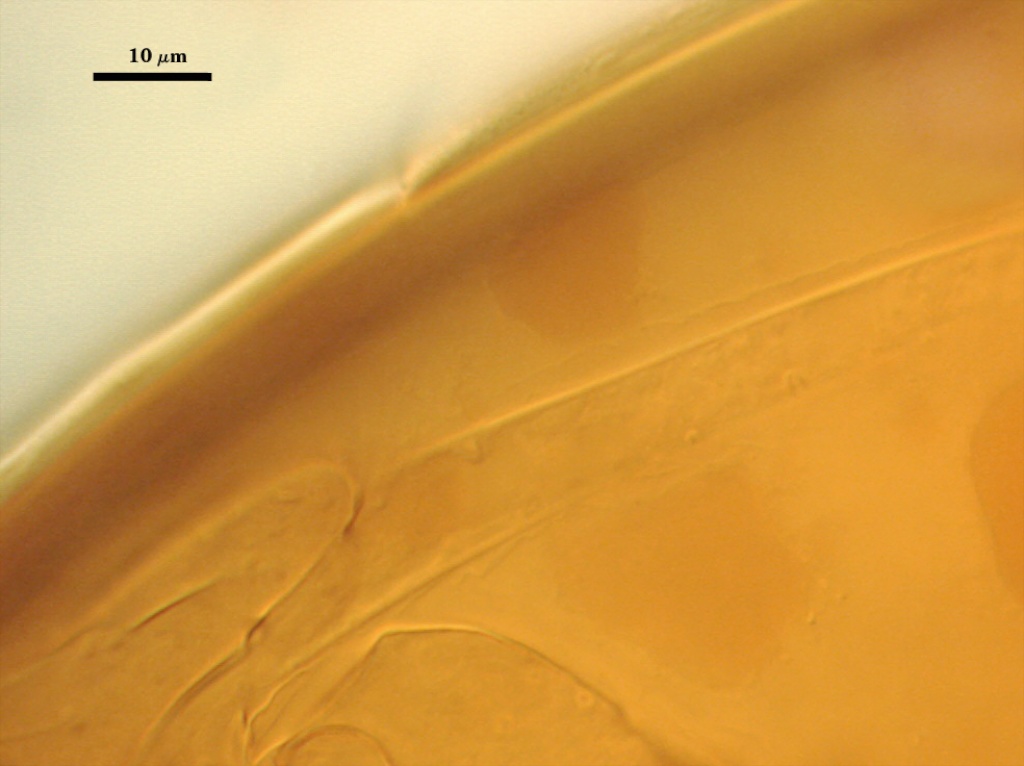
SPORE WALL: Two layers (L1 and L2) that are adherent that in juvenile spores are of equal thickness, with the laminate layer thickening as the spore wall is differentiated.
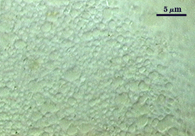
L1: An outer permanent rigid layer, yellow-brown (0-20-100-0), 0.7-1.8 m thick, tightly adherent to L2 . The surface of this layer consists of tightly packed warts less than one micrometer apart, 0.5-1.0 µm wide and 0.5-1.2 µm high.
L2: A layer consisting of pale yellow-brown (0-10-40-0) to yellow-brown (0-10-80-0) sublayers (or laminae); 6.2-8.4 µm (mean of 7.6 µm). They stain orange-brown (0-40-100-0) to red-brown (0-60-80-0) in Melzer’s reagent.
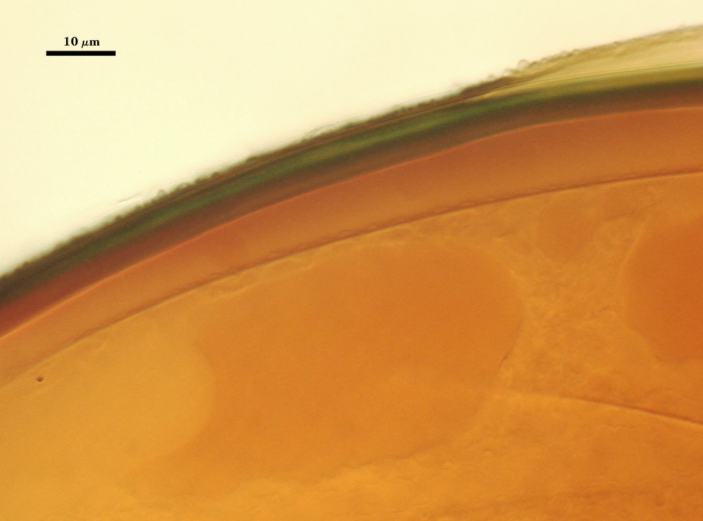
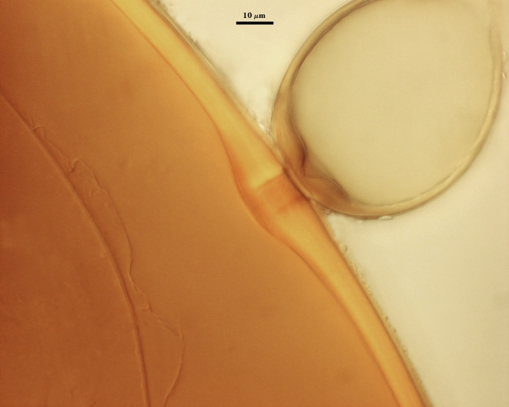
GERMINAL WALLS: One (gw1) formed independent of the spore wall and subtending hypha, consisting of two thin, often adherent layers.
| In PVLG | |
|---|---|
 |  |
| In PVLG & Melzer's reagent | |
|---|---|
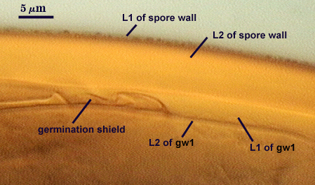 |  |
GW1: Two hyaline layers (L1 and L2) that in field-collected spores are so adherent that they appear as one layer (the condition of type specimens used to describe the species). In spores from pot cultures, the outer layer often separates in small folds from parts of the wall giving the wall a blistered appearance.
L1: A thin layer less than 0.5 µm thick and thus difficult to resolve without differential interference optics. No reaction in Melzer’s reagent.
L2: A slightly thicker layer, 0.6-1.2 µm thick. No reaction in Melzer’s reagent.
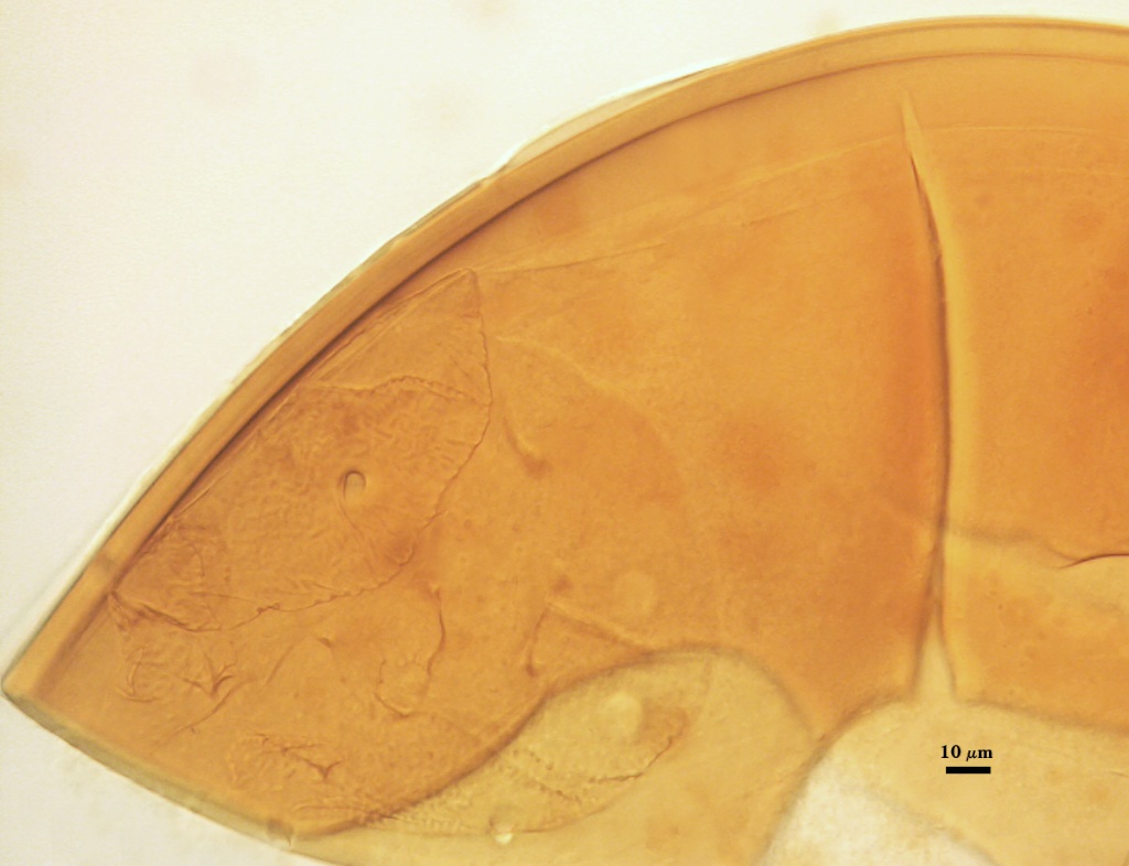
Subtending Hypha
WIDTH OF SPOROGENOUS CELL: 52-60 µm (mean = 54.9 µm)
SPOROGENOUS CELL WALL: Two hyaline layers (L1 and L2) probably are present (continuous with the two layers of the spore wall), but only L2 is readily discernible at the level of the compound microscope.
L2: Yellow-brown (0-10-80-0), 4.6-8.0 µm thick near the spore and then thinning to 1.4-1.6 µm beyond the sporogenous cell.
OCCLUSION: Closure by a plug concolorous with the laminate layer of the spore wall.
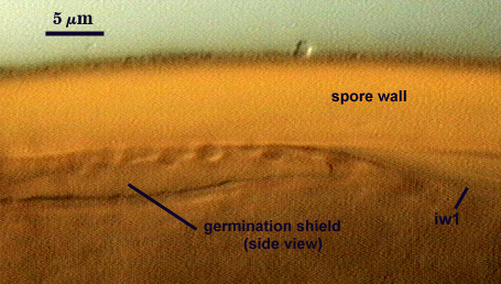
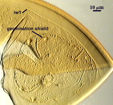
Germination
COLOR: Hyaline to pale yellow (0-10-20-0).
SHAPE: Ovoid, with length approximately 1.5 times that of the width. Margins of the shield with many convolutions that appear as deep warts when viewed from the side. It also has a rugose appearance in plan view that contribute to the warty surface in longitudinal view. Position of the shield is between the spore and germinal inner wall.
SIZE: One intact shield measured 69×114 µm.
Auxiliary Cells
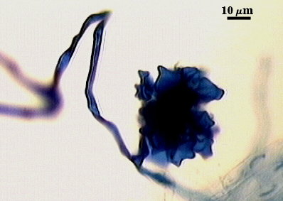
Cells in aggregates of 5-13 (mean = 8), subglobose, ovoid to clavate, borne on coiled hyaline hyphae, thin-walled (< 1 µm thick), pale cream (0-0-10-0) in transmitted light, each cell with knobby swellings 1-5 µm high and 3-10 µm wide.
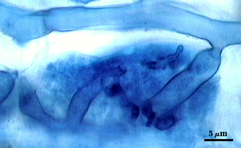
Mycorrhizae
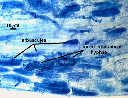
Arbuscular hyphae branch to form many fine tips from a swollen basal hypha. Intraradical hyphae 3-9 µm wide, with knobs, projections and swollen areas (up to 12 µm wide), and usually densely coiled near entry points and in outer cortical cells.
| Arbuscules in corn roots | |
|---|---|
 | 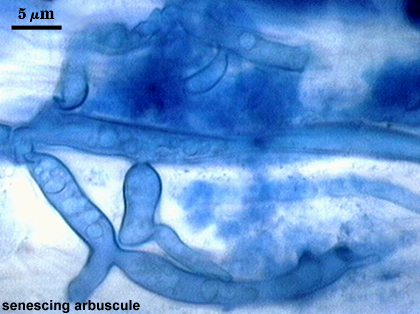 |
| Entry point and other mycorrhizal structures | ||
|---|---|---|
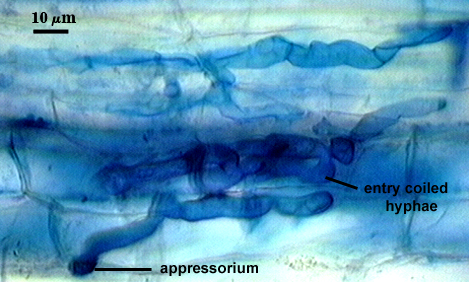 |  | 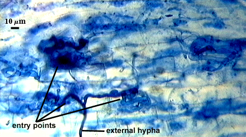 |
Extraradical hyphae of two morphological types: one wide (3-7 µm) and the other thinner (1.5-2 .0 µm). The former usually is the infective hyphae at entry points and forms knobby swellings there and near auxiliary cells. Intraradical arbuscules and hyphae consistently stain darkly in roots treated with trypan blue.
The images below can be uploaded into your browser by clicking on the thumbnail or can be downloaded to your computer by clicking on the link below each image. Please do not use these images for other than personal use without expressed permission from INVAM.
High Resolution Images | |
|---|---|
 |  |
 |  |
 |  |