Funneliformis verruculosum
(reference accession PL117B)
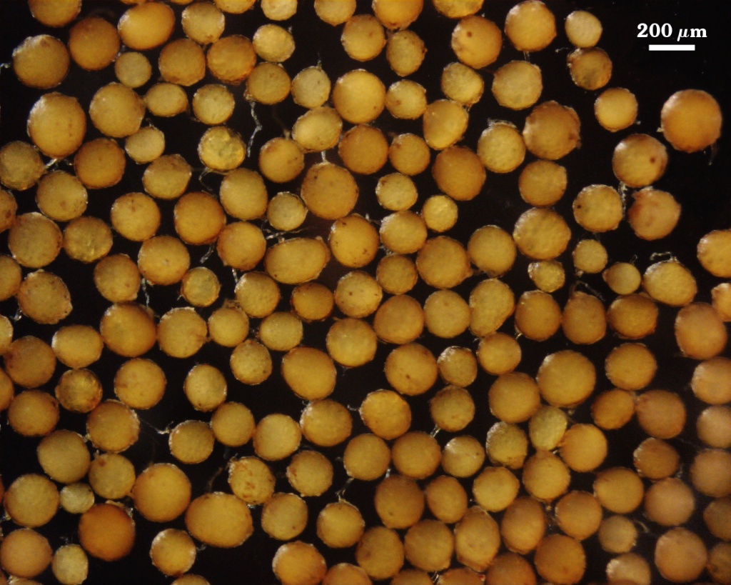
Whole Spores
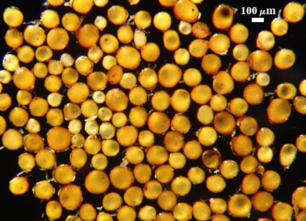
 COLOR: Bright yellowish orange (0-10-60-0) to dark brownish orange (0-40-100-0), with many bright orange (0-20-100-0)
SHAPE: Globose, subglobose, occasionally ovoid
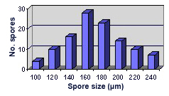
SIZE DISTRIBUTION: 100-240 µm, mean = 171 µm, n = 110
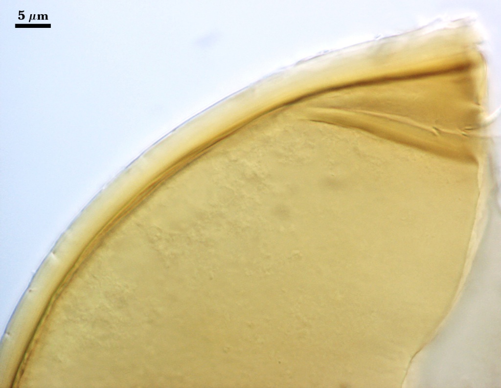
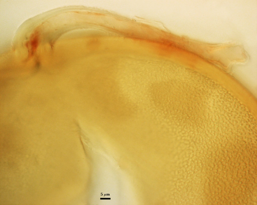
Subcellular Structure of Spores
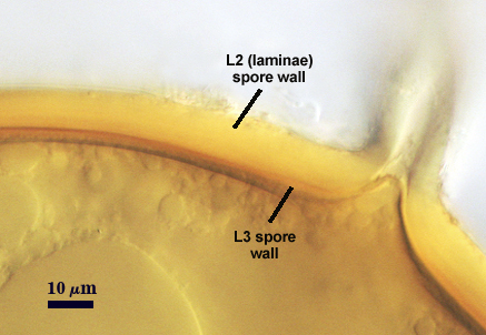
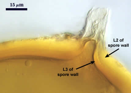
SPORE WALL: Consisting of two layers (L1 and L2) that differentiate consecutively as spores develop.
| Smashed Spores | ||
|---|---|---|
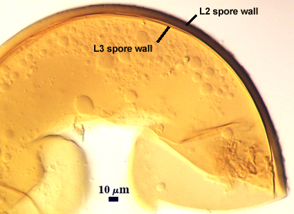 | 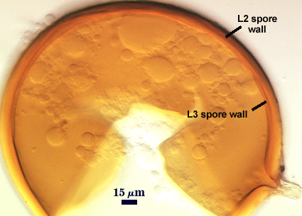 | 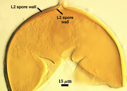 |
L1: A semiflexible, hyaline layer, 1-2 µm thick; usually sloughing and therefore absent in field-collected specimens and many spores in a mature pot culture..
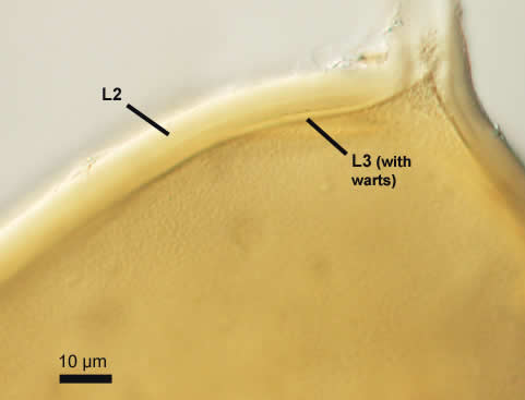
L2: A rigid layer consisting of fine adherent sublayers (or laminae), yellow to orange in color, 5-14 µm thick. Evenly distributed warts, 0.8-1.7 µm high in cross view, 0.5-0.7 µm diam in plan view, are reported by Blaszkowski and Tadych (1997) as projecting inward from the innermost sublayer, but these are seen only in a small percentage of spores in this reference culture. Given that variability, this trait is not a taxonomically informative character.
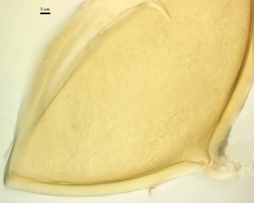
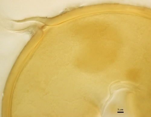
Subtending Hypha
SHAPE: Cylindrical to somewhate flared, occasionally recurved.
WIDTH: 15-28 µm (mean = 21 µm).
COMPOSITE WALL THICKNESS: 4.5-7 µm at spore base.
WALL STRUCTURE: Two layers (L1 and L2) continuous with the inner two layers of the spore wall and with the same properties. L2 extends for only 20-25 µm down length of hypha.
OCCLUSION: Recurved septum from the innermost sublayer of L2 of the spore wall.
Notes
Color of the spores is quite distinctive among glomoid species described thus far. The size range of spores in the reference culture is considerably extended at the small end than is published in the protologue (the lower limit reported to be 145 µm).
The images below can be uploaded into your browser by clicking on the thumbnail or can be downloaded to your computer by clicking on the link below each image. Please do not use these images for other than personal use without expressed permission from INVAM.
High Resolution Images | |
|---|---|
 |  |
 |  |
 |  |
Links to Gene Sequences in Genbank
Reference
- Blaszkowski, J. and M.Tadych. 1997. Glomus multiforum and G. verruculosum, two new species from Poland. Mycologia 89:804-811.