Acaulosporaceae Gerd. & Trappe
All member species form spores on or within the neck (subtending hypha) of a “sporiferous saccule.” Regardless of the position of the spore relative to the saccule hypha, spore ontogeny progressed identically. Below is the linear sequence of discrete events that start with a saccule and terminate with a fully differentiated spore having a bilayered spore wall, a an outer bilayered flexible hyaline wall (gw1), and an inner bilayered hyaline wall (gw2) on which the germination orb always develops after the spore has completed all stages.
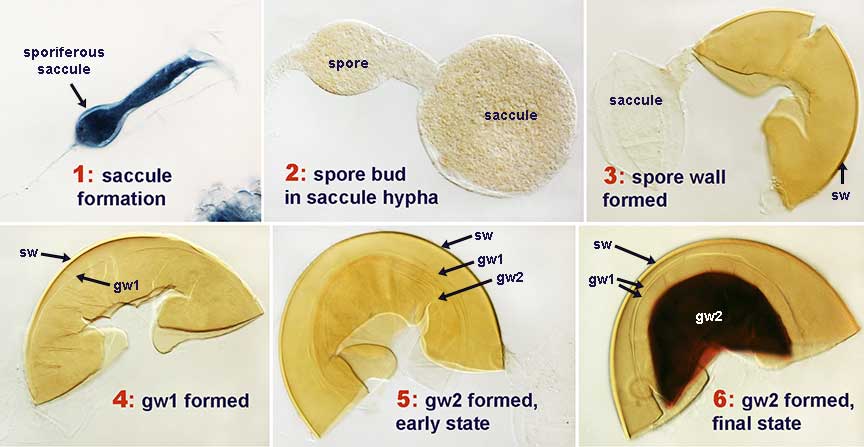
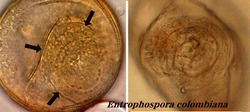
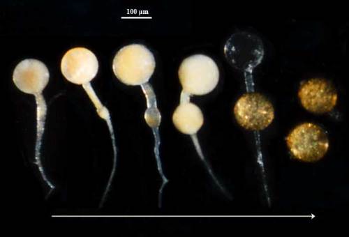
The image below shows the progression of saccule maturation concurrent with spore formation in A. colombiana (formerly Entrophospora colombiana because spore forms inside the saccule neck). The saccule forms by terminal expansion of a hypha that becomes funnel-shaped and expands to its full width before spore formation begins. The saccule differentiates a rigid saccule wall (usually 1-2 layers at most) as spore formation begins. The dense contents in the saccule begin to disperse and finally disappear as spore differentiation ends.
Entrophospora columbiana
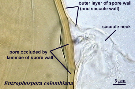
The outer layer of the spore consists of the wall of the saccule hypha (neck) and it degrades over time, sloughs, and releases the spore so that it no longer has any attachment. In other words, the spore is sessile.
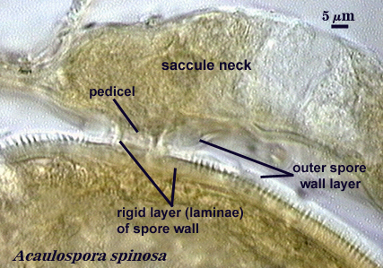
Acaulospora spinosa
The second layer (L2) of the spore wall is the one which gives structural integrity to the spore. It consists of laminae (very thin sublayers needed to provide thickness) and is the layer which varies in color and ornamentation and often is the major distinguishing feature between species. A few species have a third layer that usually is adherent but is discernible by a differential reaction in Melzer’s reagent (see Acaulospora koskei). The image here shows the laminate layer of the spore wall as well as the outer layer because all still are attached to the neck of the saccule. Note that this layer is a yellow-brown color and is ornamented on the outer surface with short densely spaced spines.
Acaulospora denticulata
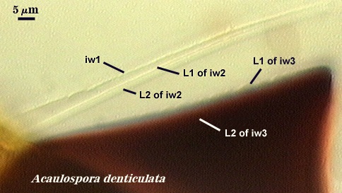
Unlike other clades with spores that have flexible inner walls (or germinal walls if you want to assign current or relict functionality), all species in Acaulosporaceae seem to have the same organization: two hyaline bilayered walls, with the inner wall serving as the base for a germination orb (producing one or more germ tubes). This germinal wall has a beaded or granular surface that is obvious just after mounting but tends to disappear as spore contents clear over time. This inner germinal wall also varies in its thickness and reaction to Melzer’s reagent. Both traits are correlated: the thicker the layer, the darker the stain reaction (pale dextrinoid to dark red purple).
Acaulospora tuberculata
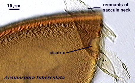
Spore contents separate from that of the subtending saccule neck by ingrowth of a “laminate” layer of the spore wall, so that the spore wall eventually shows no evidence of an opening or pore. The region of hyphal attachment usually is evident from a ridge where the spore and hyphal wall were connected and this round to ovoid scar is termed a cicatrix.
Entrophospora columbiana
A germ tube emerges from a spherical (usually) “germination orb” (so named to indicate the absence of homology with a similar structure named a “germination shield” in Scutellospora and related clades). It originates from the inner germinal wall and often is so transparent that it is hidden by oil droplets in the spore contents and is most easily observed in older spores with contents cleared.