Acaulospora koskei
(reference accession PL116)
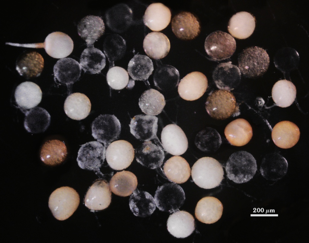
Whole Spores
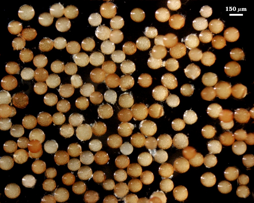
 COLOR: Pale yellow-brown (0-10-20-0) to dark orange-brown (0-60-100-10, most pale orange-brown (0-30-80-0).
SHAPE: Globose, subglobose, some oblong to irregular.
SIZE DISTRIBUTION: 120-240 µm, mean = 187 µm (n = 110).
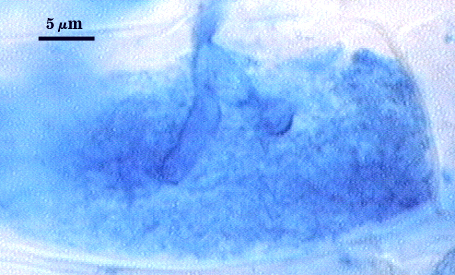
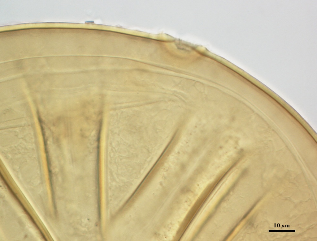
Subcellular Structure of Spores
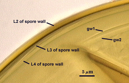
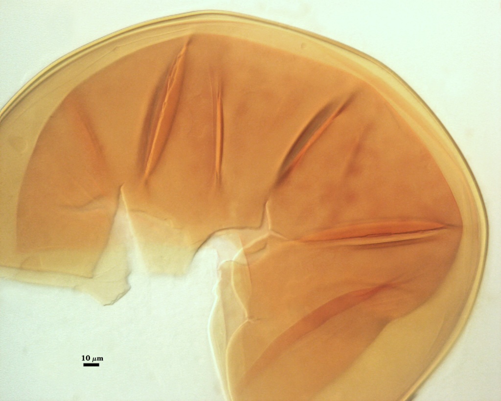
SPORE WALL: Consisting of three layers (L1, L2, and L3), the outer one continuous with the wall of the neck of the parent sporiferous saccule and inner two synthesized with expansion of the spore.
L1: Hyaline, smooth, 1.3-2.2 µm thick in juvenile spores; appears as a granular coating on the spore surface following degradation as spores age, sometimes accumulating organic debris.
L2: A layer consisting of very fine and adherent sublayers (or laminae), yellow-brown (0-10-100-0 to(0-20-100-0), 1.3-2.6 µm thick (mean of 1.9 µm). The surface of this layer is smooth.
L3: Another laminate layer concolorous with L2 in PVLG, but differentially producing an orange-brown (0-60-40-0) to darker red-brown (0-60-50-10) reaction in Melzer’s reagent when viewed through the spore wall. It is 1.2-1.6 µm thick under these conditions. At maturity, the pore between spore and saccule neck is closed by both L2 and L3 and thus functions like an “endospore”.
| Spores in PVLG. Far left photo of spore in sodium azide 1yr. | ||
|---|---|---|
|
|
|
| Others freshly extracted. All spores freshly extracted and mounted in 1:1 v/v PVLG and Melzer’s reagent | ||
|---|---|---|
|
|
|
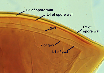

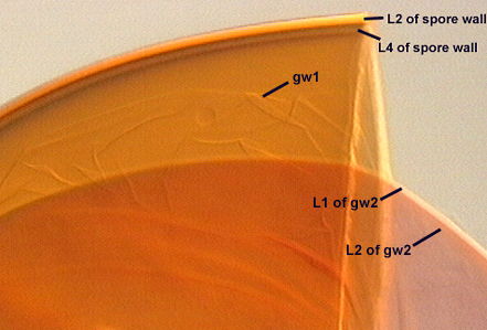
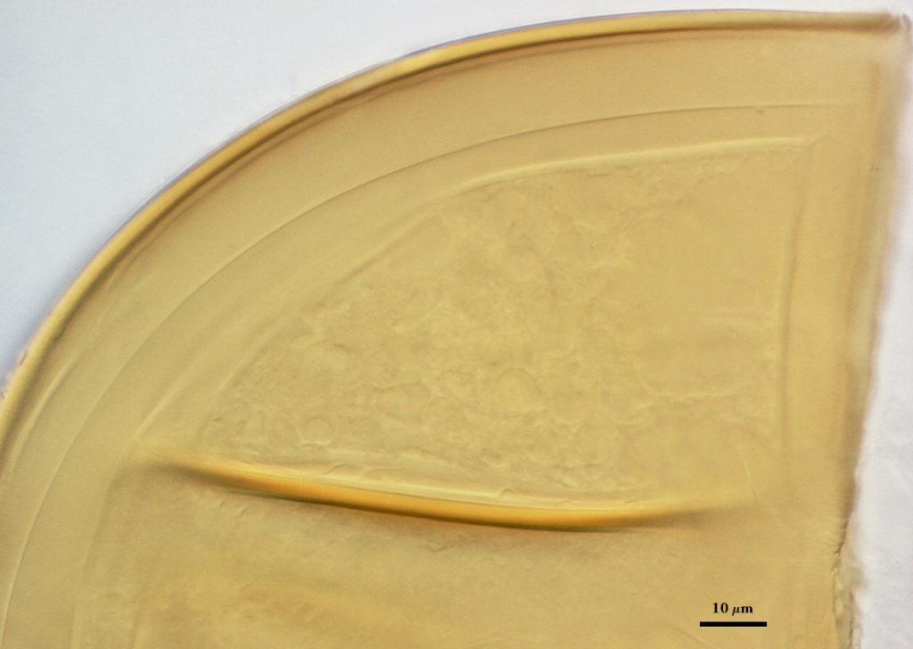
GERMINAL WALLS: Two flexible hyaline inner walls (gw1 and gw2) are present..
GW1: Appearing to consist of a single hyaline layer, 0.7-1.0 µm thick, but some spores suggest that this wall may be bi-layered, with each layer of equal thickness. If these layers are so adherent and thin, then resolving both layers would be difficult at the light microscope level.
GW2: Two adherent layers (L1 and L2) are formed. L1 is 0.6-1.0 µm thick and coated on the surface with granular excresences (or “beads”) that tend to become dislodged and float away when pressure is applied to it. These “beads” often disappear in common mountants within several weeks after placement on a slide (see photos above), but are quite obvious in spores stored for longer than several months in common preservatives like formalin or sodium azide (see left photo in first row above). L2 is 0.5-1.1 µm thick and stains a light reddish-purple (20-60-20-0) in Melzer’s reagent.
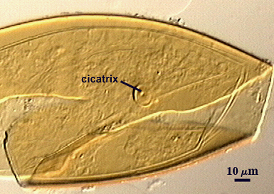
Cicatrix
Circular to ovoid scar indicating region of contact between spore and saccule neck during spore synthesis; 13-16 µm in diameter (mean = 14.2 µm).
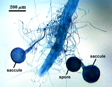
Sporiferous Saccule
COLOR: Hyaline
SHAPE: Mostly globose.
SIZE: 140-180 µm, mean = 162 µm.
SACCULE WALL: One layer, smooth surface, 1.25-4.2 µm thick (mean = 3.2 µm).
DISTANCE 50-100 µm (mean = 78 µm).
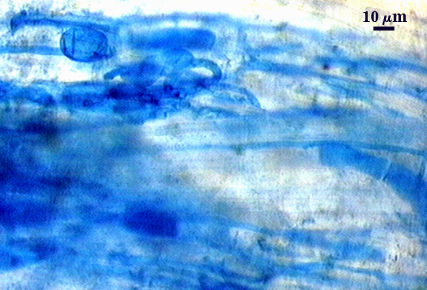
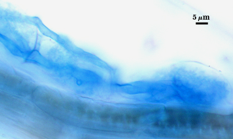
Mycorrhizae
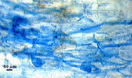
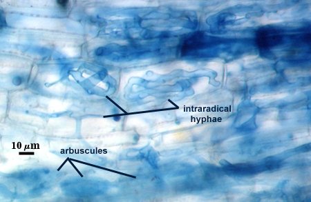
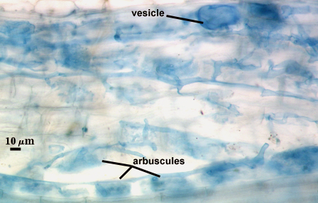
Arbuscules usually stain faintly in trypan blue and often are hard to see, although the intensity of staining varies considerably within and between roots. Oblong to irregular vesicles and attached hyphae often cluster within roots; they tend to stain more darkly and thus are more readily detected and measured. Intraradical hyphae stain with variable intensity, usually darkest near entry points and lighter near the outer edges of “infection units”. Hyphae are widest near entry points (4-6 µm) and often exhibit some coiling. They become narrower (1.5-3 µm) as they grow into the root cortex, extending parallel with the root axis and with neighboring hyphae. Interconnections between hypha are numerous, with branches from acute to oblique angles.
| Arbuscules in cortical cells of corn root | ||
|---|---|---|
|
|
|
| All mycorrhizae structures in corn | |||
|---|---|---|---|
|
|
|
|

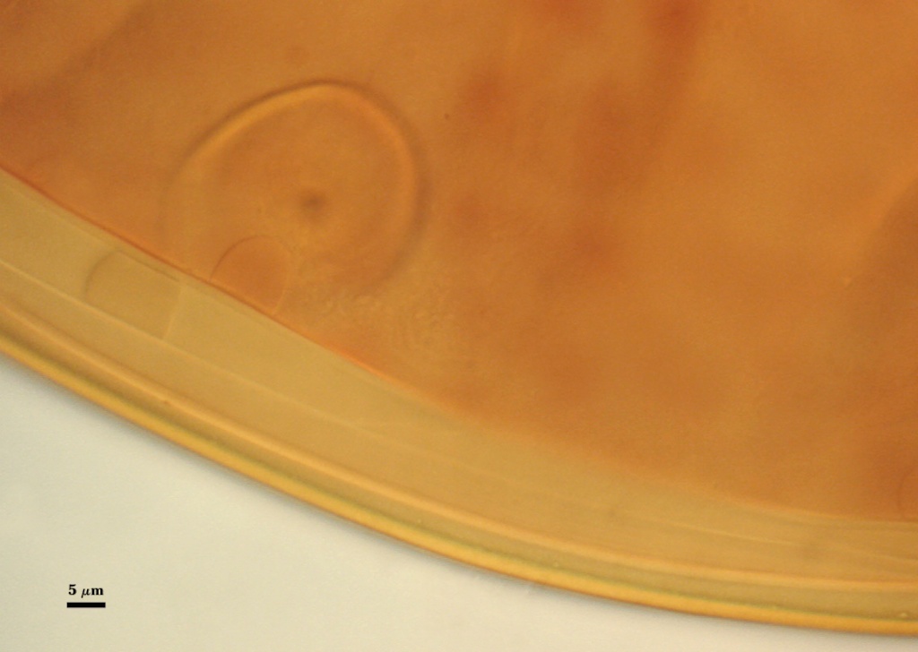
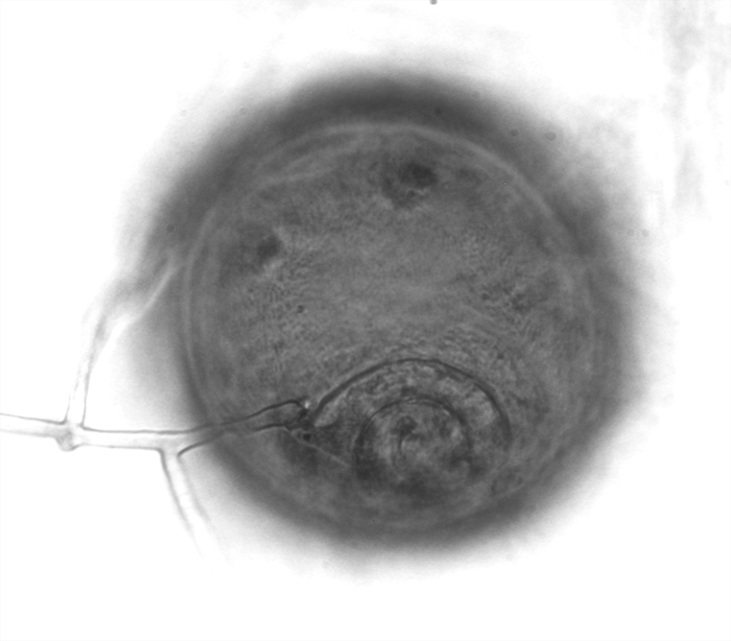
Germination
An ovoid “germination orb” forms on gw2, from which germ tubes form and penetrate through the spore wall. This orb is difficult to see except in older spores where contents have cleared with fusion of lipid globules in the spore lumen.
Notes
This species closely resembles A. laevis in size and color; differing mostly in that it produces a reaction to Melzer’s reagent in L3 of the spore wall (not reactive in spores of A. laevis) and L2 of gw2 (not reactive or only faintly reactive in spores of A. laevis). Under a dissecting microscope, these two species are indistinguishable.
High Resolution Images | |||
|---|---|---|---|
 |  | ||
 |  | ||
 |  | ||
 | |||