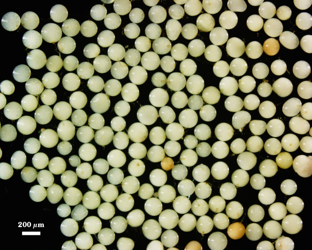
Gigaspora albida
(reference accession BR214)
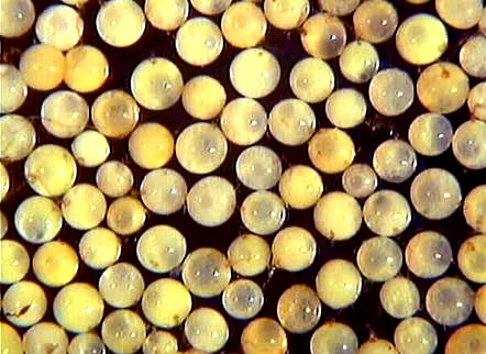
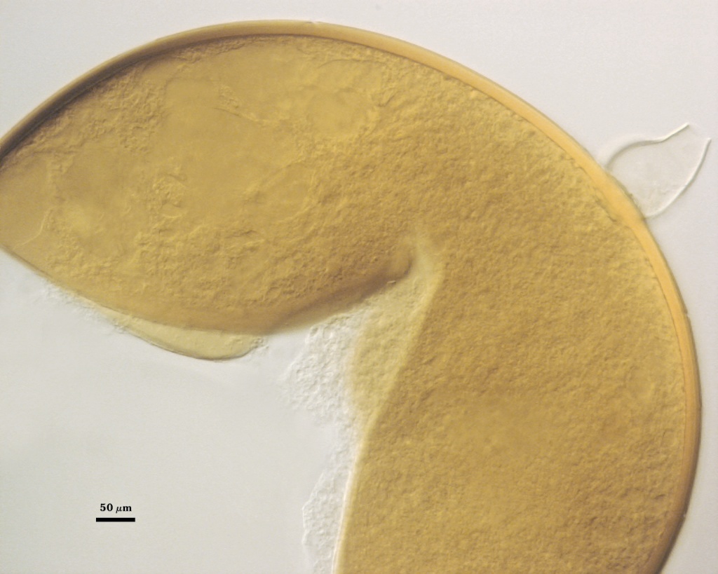
Whole Spores
 COLOR: Cream with pale green tint (0/0/15/0 to 5/0/20/0).
SHAPE: Globose to subglobose.
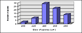
SIZE DISTRIBUTION: 200 – 280 µm, mean = 250 µm.
Subcellular Structure of Spores
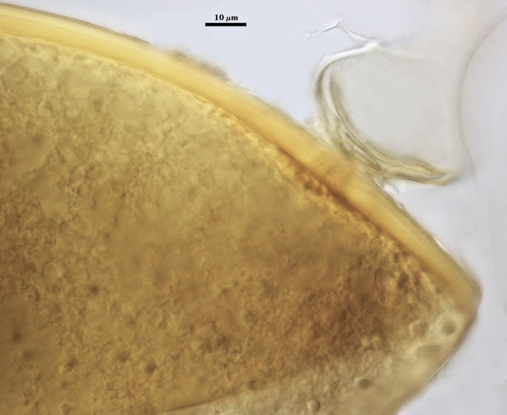
SPORE WALL: Three layers (L1, L2, and L3), the first two adherent and of equal thickness in juvenile spores, with L2 thickening as the spore wall is differentiated. L3 differentiates as a prelude to germ tube formation.
| Gigaspora albida | |
|---|---|
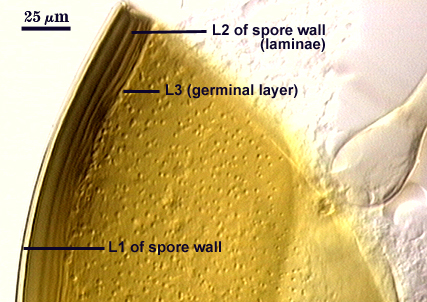 |  |
L1: An outer permanent rigid layer with a smooth surface, hyaline to pale yellow (0-0-10-0), 2.0-3.2 m thick.
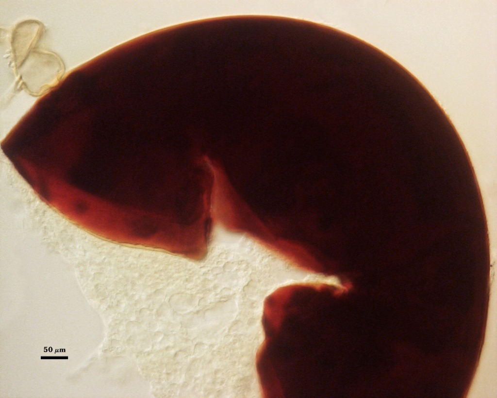
L2: A semi-plastic layer consisting of yellow (0-10-60-0) to brownish yellow (0-10-100-0) sublayers (or laminae) that increase in number with differentiation; varying considerably in thickness in mature spores; 14-26 m thick (15-25 µm in the same spore). This variation is attributed to plasticity of sublayers that swell and spread with applied. Sublayers are pale brown (0-10-60-0) in PVLG, staining dark red-brown (20-80-100-0) to a very dark red-purple (60-80-70-10) in Melzer’s reagent.
L3: A “germinal” layer that is concolorous with theL2 layer and is adherent. This layer is distinct only at the ultrastructural level, where it appears electron dense. Numerous “warts” or “papillae” form on the inner surface of this layer, and they are especially concentrated in regions where germ tubes form (usually in close proximity to the suspensor cell);warts 1.6-4 µm high in germinating spores.
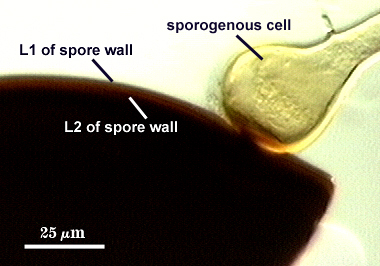
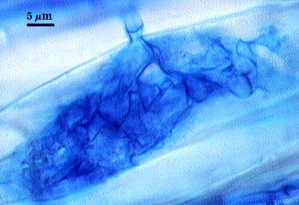
Subtending Hypha
WIDTH OF SPOROGENOUS CELL: 32-45 µm (mean = 41 µm) (see photos above).
SPOROGENOUS CELL WALL: Two hyaline layers (L1 and L2) probably are present (continuous with the first two layers of the spore wall), but only L2 is easily discernible with observation at the level of the compound microscope.
L2: Brownish yellow (0-10-60-0), 2.5-4.2 µm thick near the spore and then thinning to 1.2-1.4 µm beyond the sporogenous cell.
OCCLUSION: Closure by a plug concolorous with L2 of the spore wall.
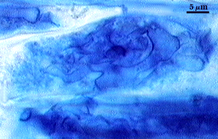
Germination
Germ tube forms in vicinity of warty protruberances on inner surface of L3 of the spore wall, 16-18 µm wide after emergence from the spore; the germ tube holes are 8-13 µm wide.
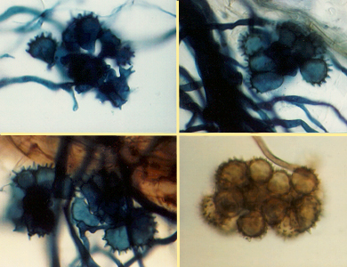
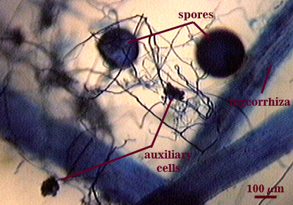
Auxiliary Cells
Cells in aggregates of 4-20, subglobose to ovoid to clavate, borne on tightly coiled hyaline hyphae, thin-walled (< 1 µm thick), hyaline to pale cream (0-0-10-0); each cell with narrow projections 1.5-2.0 µm wide and 2.0-10.0 µm high.
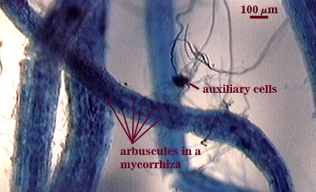
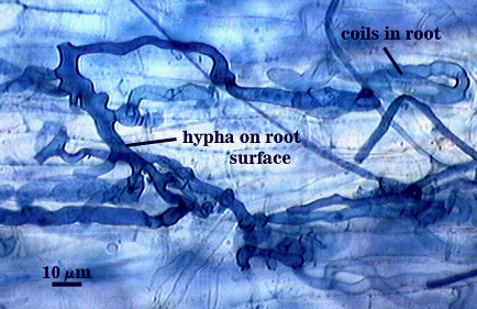
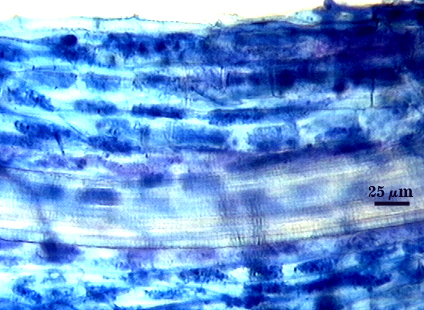
Mycorrhizae
Intraradical arbuscules and hyphae consistently stain darkly in roots treated with trypan blue. Arbuscules produce fine-branches from a swollen basal hypha(e) that are easiest to see as tips degrade. Intraradical hyphae 3-8 µm in diameter, with inflated areas up to 10 µm and knob-like projections distributed along length, usually densely coiled near entry points.
| Arbuscules in cortical cells of corn roots | |
|---|---|
|
|
| Views of trypan blue-stained corn roots under a dissecting microscope | |
|---|---|
|
|
| External hyphae, entry points, and arbuscules in corn mycorrhizae | |
|---|---|
|
|
Notes
Immature spores often are salmon-colored with a slight pink tint (0-10-20-0 to 0-20-60-0). L1 and L2 of the spore wall in juvenile spores are equally 1.6- 2 µm thick. Spores collected from the field can be dark brown to black from parasitism and colonization of the spore lumen by a wide range of soil microorganisms (especially actinomycetes and other saprobic fungi). This species often is confused with Gi. gigantea in the field because both can become brown with a green tint when old, parasitized, or dead.
The images below can be uploaded into your browser by clicking on the thumbnail or can be downloaded to your computer by clicking on the link below each image. Please do not use these images for other than personal use without expressed permission from INVAM.
High Resolution Images | |
|---|---|
 |  |
 |  |