Gigaspora gigantea
(reference accession MA435A)
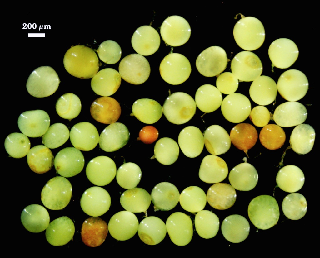
Whole Spores
| Gigantea sproes | ||
|---|---|---|
|
|
|
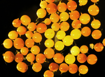
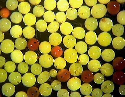
 COLOR: Bright greenish yellow (10-0-100-0) to bright yellow-green (20-0-100-0).
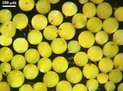
SHAPE: Globose to subglobose, rarely irregular.
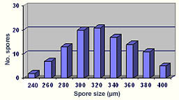
SIZE DISTRIBUTION: 240-400 µm, mean = 324µm (n = 110).
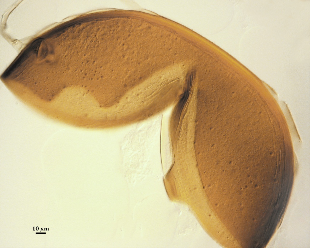
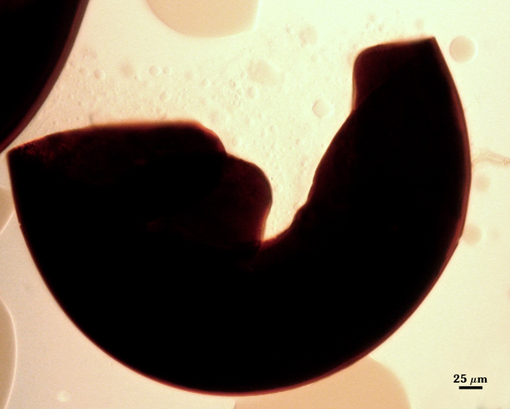
Subcellular Structure of Spores
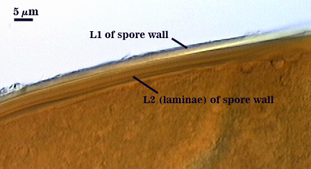
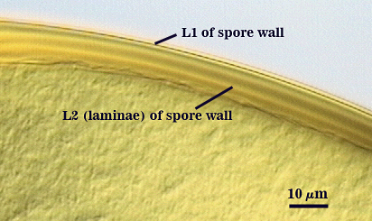
SPORE WALL: Three layers (L1, L2, and L3), the first two adherent and of equal thickness in juvenile spores, with L2 thickening as the spore wall is differentiated and L3 differentiating as a prelude to germ tube formation.
| Spores in PVLG | |
|---|---|
|
|
| Spores in 1:1 v/v | |
|---|---|
|
|
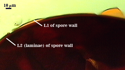
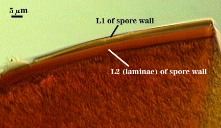
L1:* An outer permanent rigid layer, pale yellow (0-0-20-0), 2.8-3.6 m thick. The surface is smooth, although debris may be attached in older spores, especially those collected from field soils.
L2: A layer consisting of sublayers (laminae) that increase in number with thickness due to plasticity that causes swelling and spreading of individual sublayers; 8-29 m (mean = 16.9 µm) thick; appearing thinnest in older spores (8-14 µm). Color ranges from yellow (0-10-80-0) to brownish-yellow (0-10-100-0) in PVLG, staining dark red-brown (20-80-100-10) to very dark red-purple (60-80-70-10) in Melzer’s reagent.
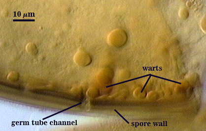
L3: A “germinal” layer that is concolorous and adherent with L2 (a laminate layer). This layer usually can be resolved only at the ultrastructural level, where it appears electron dense. Numerous “warts” or “papillae” form on the inner surface of this layer, and they are especially concentrated in regions where germ tubes form (usually in close proximity to the suspensor cell); warts 1.6-5 µm high in germinating spores and 2-3 µm wide.
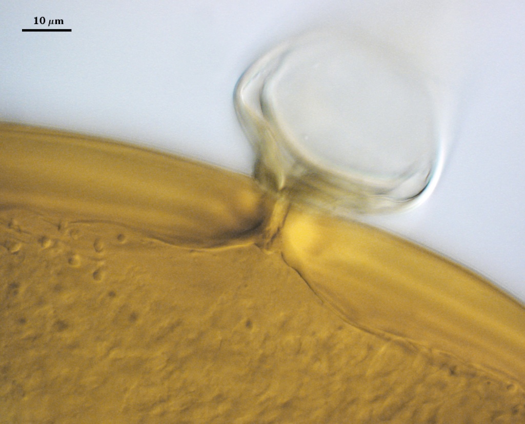
Subtending Hypha
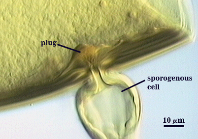
WIDTH OF SPOROGENOUS CELL: 38-54 µm (mean = 47.8 µm)/
SPOROGENOUS CELL WALL: Two hyaline layers (L1 and L2) probably are present (continuous with the first two layers of the spore wall), but only L2 is readily discernible at the level of the compound microscope.
L2: Brownish yellow (0-10-60-0), 2.0-6.4 µm thick near the spore and then thinning to 1.2-1.6 µm beyond the sporogenous cell.
OCCLUSION: Closure by a plug concolorous with the laminate layer of the spore wall.
Germination
Germ tube forms in vicinity of warty protruberances on inner surface of L3 of the spore wall, 12-16 µm wide after emergence from the spore; the germ tube holes are 6-10 µm wide.
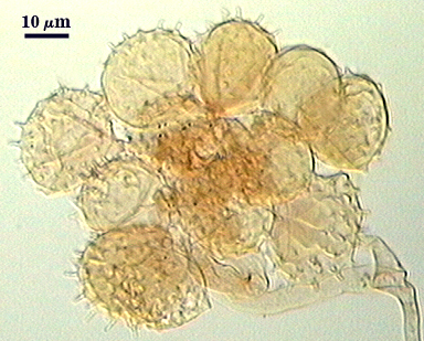
Auxiliary Cells
Cells in aggregates of 4-20, subglobose to ovoid to clavate, borne on tightly coiled hyaline hyphae, thin-walled (<1 m thick), hyaline to pale cream (0-0-10-0); each cell with narrow projections (1.5-2.0 wide and 2.0-10.0 high).
<>
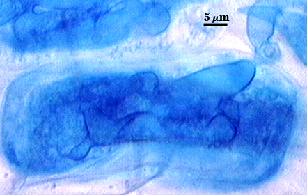
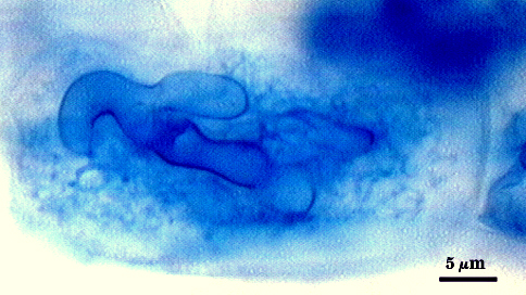
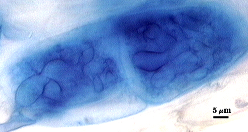
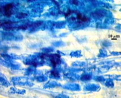
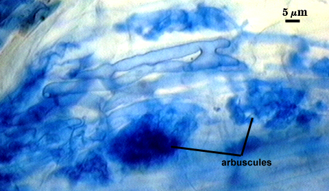
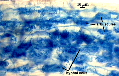
Mycorrhizae
Arbuscules and hyphae in cortical cells of sorghum, sudangrass, corn and red clover roots stain darkly in trypan blue. Numerous vesicles (or spores) often form near entry points along with the arbuscule-hyphal network. It is uncertain as the extent intraradical vesicles are still able to further differentiate into spores, because they can co-occur in roots.
| Arbuscule in cortical cell of a corn root | ||
|---|---|---|
|
|
|
| Mycorrhizae in corn roots | ||
|---|---|---|
|
|
|
Notes
Immature spores often are salmon-colored (0-20-60-0) to orange-brown (0-40-80-0) with opaque contents. Old senescing spores also turn orange-brown, but the contents are not opaque. Color of spores is conferred by pigmentation in the spore contents rather than the spore wall, which might account for the dramatic shift from orange-brown to green with maturation (or deterioration). Some spores collected from the field appear leached out, and thus can be confused with Gi. albida at the small end of the size range.
Mature spores were described by Nicolson and Gerdemann (1968) as being as large as 812 µm, but they rarely exceed 450 µm in cultures of INVAM isolates.
The images below can be uploaded into your browser by clicking on the thumbnail or can be downloaded to your computer by clicking on the link below each image. Please do not use these images for other than personal use without expressed permission from INVAM.
High Resolution Images | |
|---|---|
 |  |
 |  |
Reference
- Nicolson, T. H. and J. W. Gerdemann. 1968. Mycorrhizal Endogone species. Mycologia 60:313-325.