Scutellospora biornata
Voucher Specimens
This description is a combination of information obtained from the protologue (Spain et al., 1989), voucher specimens prepared from a vial of living spores contributed by Joyce Spain, and universal patterns of morphological organization and structure in Gigasporaceae. A living culture of this species has never been successfully established by INVAM.
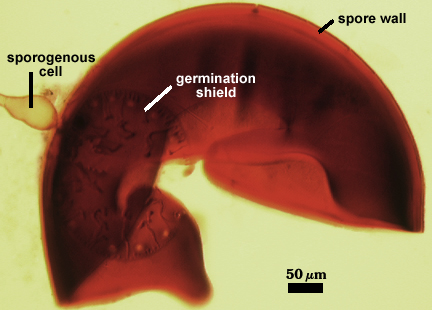
Spores are borne singly in soil on a bulbous sporogenous cell characteristic of the genus. They are globose to subglobose, (120) 260-450 (493) µm diam, and yellowish to orange-brown in color.
| Spore wall in Melzer’s reagent | Plasticity of L2 of the spore wall |
|---|---|
 |  |
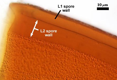
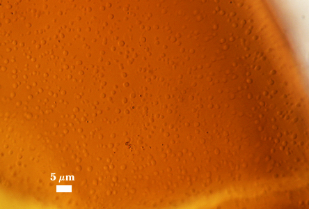
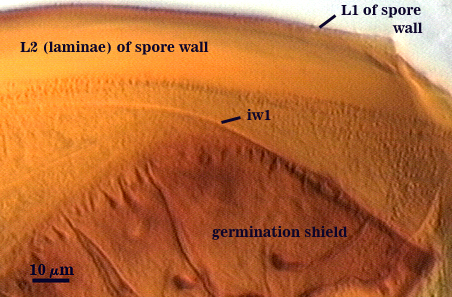
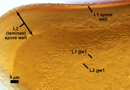
The spore wall is reported (Spain et al., 1989) to consists of three layers, but whenever detected more than two (L1 and L2). The outermost layer (L1) is brown, 0.5-1 µm thick, with blunt tapering projections on the surface that are (0.5) 1-3 µm wide at the base and up to 2 µm high. The inner layer (L2) is pale orange-brown, with numerous finely adherent sublayers (or laminae). This layer swells in acidic mountants and can exhibit considerable plasticity. This property consistently is associated with a dark red-brown to red-purple reaction in Melzer’s reagent (much like that in germinal walls of other Scutellospora species, such as S. pellucida). With only light crushing, L2 is 5-10 µm thick, but when pressure is applied more heavily or repeatedly, this layer can spread to almost 25 µm thick (see photo at right). Sublayers then appear oddly distributed and the innermost surface can produce numerous fine folds suggestive of another layer. Complicating interpretation of L2 is the presence of crowded blunt projections 0.5-1 µm wide at the base and up to 2 µm high on the inner surface.
| Spore wall properties in PVLG | ||
|---|---|---|
 |  |  |
One germinal wall is present, described by Spain et al. (1989) as having three layers. However, we observed only the bi-layered pattern common to all other members of the genus. This germinal wall is hyaline, flexible, and of two layers (L1 and L2). L1 is 0.5-1 µm thick, with a beaded surface, and nonreactive in Melzer’s reagent. L2 is 1-2 µm thick and produces a pale pink to light reddish-brown (dextrinoid) reaction in Melzer’s reagent.
| Germinal wall properties in PVLG | ||
|---|---|---|
 |  | 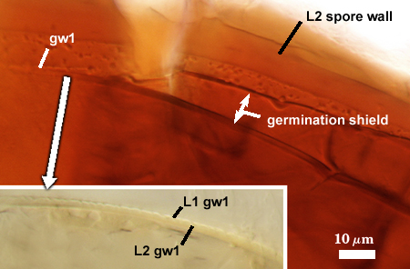 |
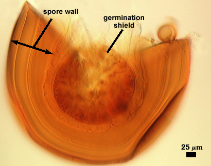
The germination shield is brown, ovoid to slightly ellipsoid, (113) 188-238 x (113) 188-275 µm, forming on the germinal wall, producing single to multiple germ tubes (see photo at top of page and below).
The sporogenous cell is brown (30) 50-60 (65) µm wide. The cell wall is a continuation of the layers of the spore wall, 2-4 µm thick at the spore base. with warty projections wider than those of the spore wall.
Auxiliary cells are brown, knobby, 32-48×37-48 µm diam., forming in clusters of 10-20 on coiled, thin-walled (< 1 µm) brown coenocytic hyphae 3-5 µm wide.
Mycorrhizae stain darkly in trypan blue. Arbuscules appear to be typical of the genus, with numerous coiled hyphae in infection units.
Reference
- Spain, J. L., E. Sieverding, and S. Toro T. 1989. Scutellospora biornata: A new species in the Endogonaceae from the Llanos Orientales of Colombia. Mycotaxon 35:219-227.