Acaulospora bireticulata
Voucher Specimens
= A. elegans (?)
Subcellular Structure of Spores
This description is a combination of information obtained from the protologue (Rothwell and Trappe, 1979), type specimens, and universal patterns of morphological organization and structure in Acaulosporaceae. A living culture of this species has never been obtained by INVAM.
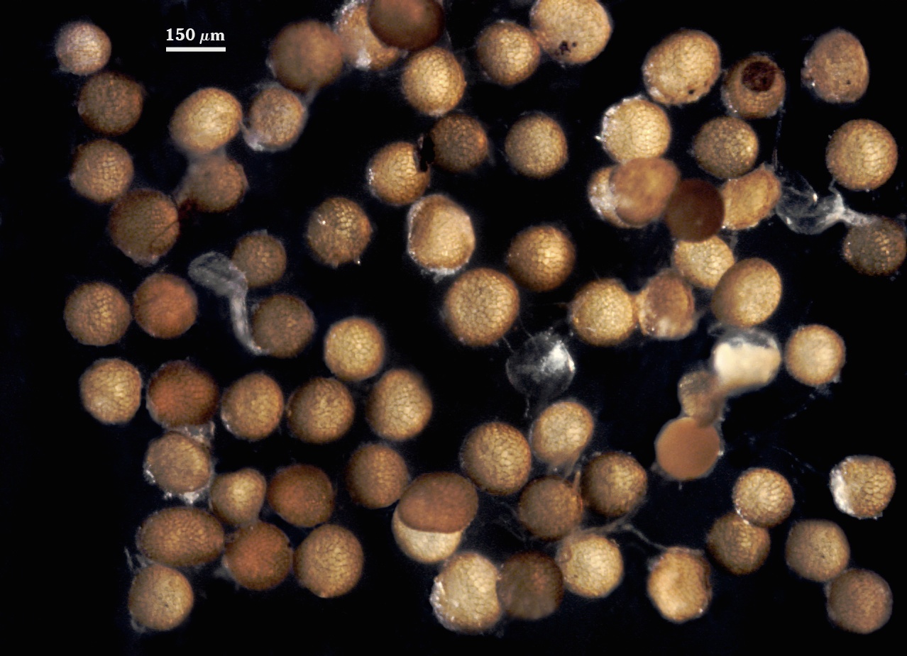
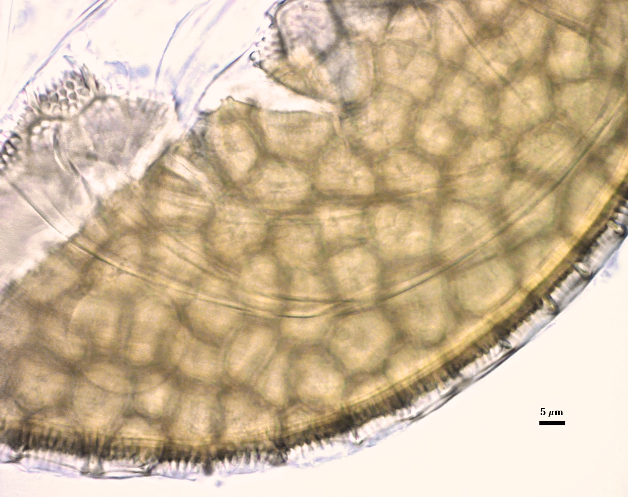
Spores borne laterally from the neck of a sporiferous saccule; singly in soil. They are globose, 150-155 µm diam, subhyaline in youth, becoming light brown by maturity.
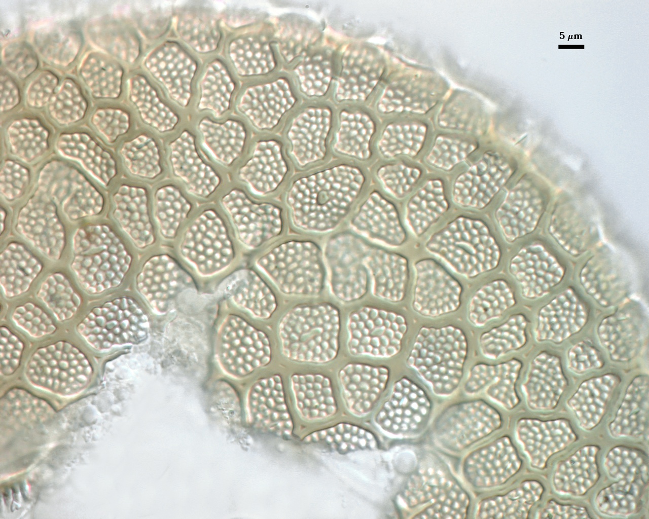
SPORE WALL: Three layers (L1, L2 and L3). The outer layer (L1) is not described in the protologue nor is it in evidence in the type specimens. However, it is assumed to develop as a continuation of the wall of the hypha subtending the sporiferous saccule, is hyaline, and sloughs with loss of the saccule. Thickness is unknown. L2 is the rigid laminate layer, differentiating “a polygonal reticulum, the ridges 1.5-2×2 µm with sinuous, dark grayish-green sides. The depressed central stratum is paler; ridges occasionally branched toward the center of polygons or forming irregular, isolated projections at polygon centers. Polygons 6-18 um long, the enclosed spore surface beset with round-tipped, 4- to 6-sided processes 1×1 µm to give the appearance of an inverted reticulum.” L3 is a thin layer which may be concolorous with the laminate layer of the spore wall, but it appears hyaline because of its thinness. This layer is easily mistaken for a germinal wall because it separates readily from L2, but it also is a structure invariant among species of Acaulospora (though apparently absent in Entrophospora).
| Spore subcellular structure (in Melzer's reagent) | |
|---|---|
|
|
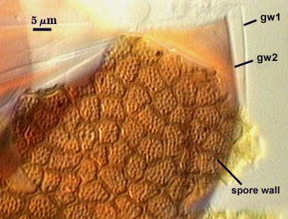
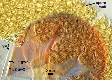
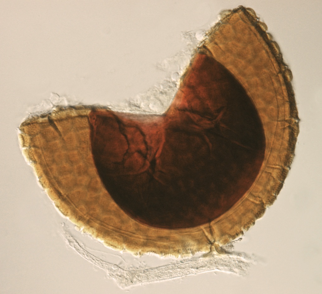
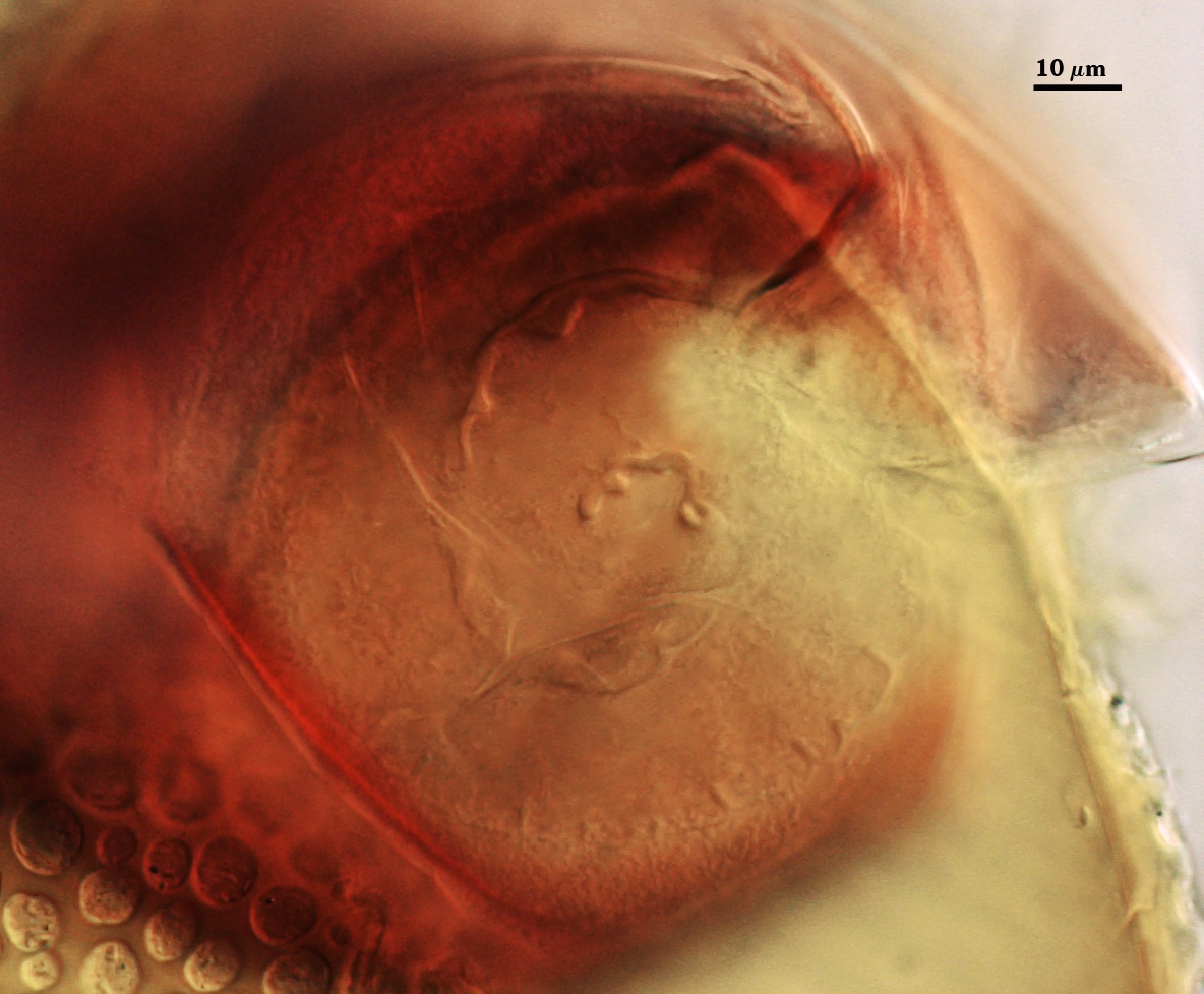
GERMINAL WALLS: Rothwell and Trappe are not clear about their description of inner wall structure, writing “spore walls of three layers, each about 1 µm thick, the outer layer dark grayish green and the inner layers hyaline”. The grayish green layer is presumed to be L3 of the spore wall. The hyaline layers are considered the two germinal walls common to all Acaulosporaceae (gw1 and gw2 in photos above).
GW1: Probably bi-layered, but hard to tell in preserved specimens (fixatives tend to tightly bind layers together). If bi-layered, both layers are hyaline, flexible, and probably of near-equal thickness.
GW2: wo hyaline layers, the outer layer “beaded” and thinner than the inner layer, which produces a red-brown reaction in Melzer’s reagent. Given that this layer does not appear thick (at least 4-5 µm) with fixation, then it is likely that the phenotype observed is true to healthy spores and not an artefact of preservation/fixation.
The sporiferous saccule is globose to subglobose and 127-135 µm diam. The saccule neck is thin-walled, tapering from 10-30 µm at the saccule base to 2.5-7.5 um. Color of the walls of the saccule or neck hypha are not mentioned.
This species appears to be identical to A. elegans except for pattern of ornamentation in the laminate layer (L2 of the spore wall). Rothwell and Trappe describe that of A. elegans as being a single-layered reticulum over crowded spines only 0.5 µm high and that of A. bireticulata as being a 3-layered reticulum over “angular processes” 1 µm high. In reality, both phenotypes can be seen in spores of each species, depending on plane of focus and depth of field. Therefore, it is likely that these two species are synonymous.
High Resolution Images | ||
|---|---|---|
 |  | |
 |  | |
 | ||
Reference
- Rothwell, F. M. and J. M. Trappe. 1979. Acaulospora bireticulata sp. nov. Mycotaxon 8:471-475.