Acaulospora elegans
Voucher Specimens
= A. bireticulata (?)
Subcellular Structure of Spores
This description is a combination of information obtained from the protologue (Gerdemann and Trappe, 1974), type specimens, and universal patterns of morphological organization and structure in Acaulosporaceae. A living culture of this species has never been obtained by INVAM.
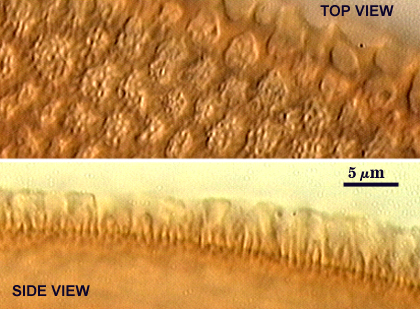
Spores forming singly in soil, borne laterally from the neck of a sporiferous saccule. They are 140-285×145-330 µm diam, globose to subglobose or ellipsoid to reniform, dark brown, with a dull surface due to light scattering from surface ornamentation.
SPORE WALL: Consists of two layers (L1 and L2). The outer layer (L1) was not reported by Gerdemann and Trappe (1974) nor was it observed in the type specimens examined. It is assumed to by formed, because it is the layer continuous with the wall of the hyphal neck subtending the saccule and is found universally in all cultured species of Acaulosporaceae. It also is assumed to be hyaline and sloughing with loss of the saccule. Layer thickness is unknown. L2 is better characterized, consisting of laminae within which are differentiated crowded, light brown spines 2 µm wide and 0.5 µm high,with total layer as much as 12 µm. An alveolate reticulum of hyaline ridges 1×5-6 µm is superimposed on the spines, with the reticulum fragmented on some spores. Alveoli are 4-8 µm long.
| Surface ornamentations |
|---|
|
| Spore subcellular structure (in Melzer's reagent) | ||
|---|---|---|
|
|
|
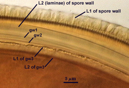
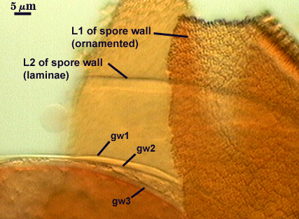
GERMINAL WALLS: Gerdemann and Trappe describe “three hyaline walls which total up to 15 µm thick”. Type specimens indicate an organization of two bi-layered germinal walls (gw1 and gw2, labelled in photos above) like other Acaulosporaceae.
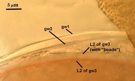
GW1: Two thin and adherent hyaline flexible layers, which appear to be of near-equal thickness; not reactive in Melzer’s reagent.
GW2: Two hyaline layers, the outer layer “beaded” and thinner than the inner layer, which produces a red-brown reaction in Melzer’s reagent. Given that this layer does not appear thick (at least 4-5 µm) with fixation, then it is likely that the phenotype observed is true to healthy spores and not an artefact of preservation/fixation.
The sporiferous saccule is globose to ellipsoid, 150-240 µm diam with a pale brown wall 1-3 µm thick, becoming empty and shrunken and then detaching at spore maturity. The hyphal neck is 40-60 µm wide with a pale brown wall 1-3 µm wide.
This species appears to be identical to A. bireticulata except for pattern of ornamentation in the laminate layer (L2 of the spore wall). Gerdemann and Trappe (1974) describe the spore wall of A. bireticulata as being a 3-layered reticulum over “angular processes” 1 µm high, whereas the spore wall of A. elegans is described as being a single-layered reticulum over crowded spines only 0.5 µm high. In reality, both phenotypes can be seen in spores of each species, depending on plane of focus and depth of field. Therefore, it is likely that these two species are synonymous.
Reference
- Gerdemann, J. W. and J. M. Trappe. 1974. Endogonaceae in the Pacific Northwest. Mycologia Mem. 5:1-76.