Paraglomus brasilianum
(reference accession BR105)
Whole Spores
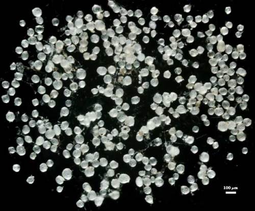
 COLOR: Subhyaline (0-0-0-0) to cream (0-0-5-0).
SHAPE: Globose to subglobose, some irregular.
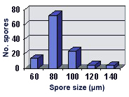
SIZE DISTRIBUTION: 60-140 µm, mean = 85 µm (n = 120).
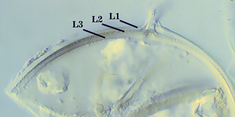
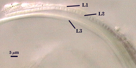
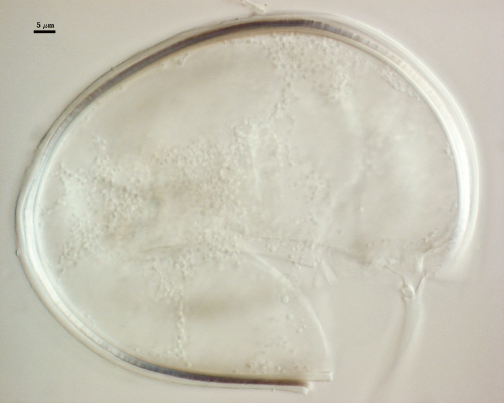
Subcellular Structure of Spores
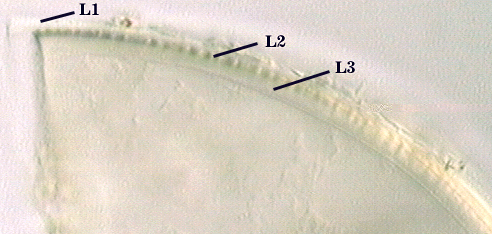
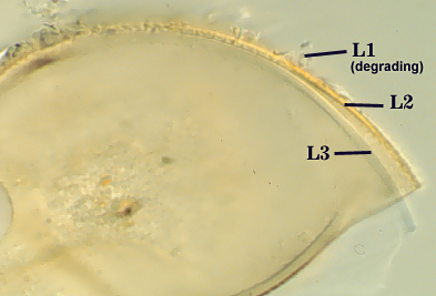
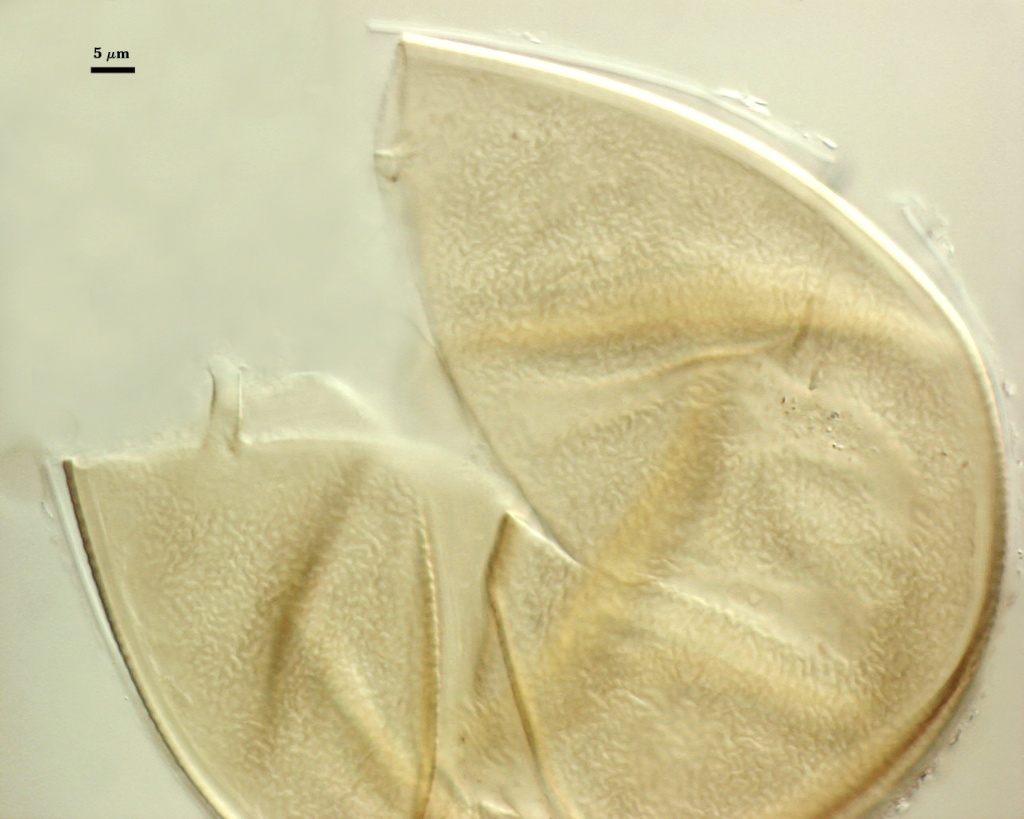
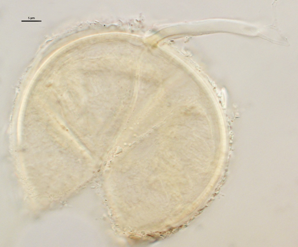
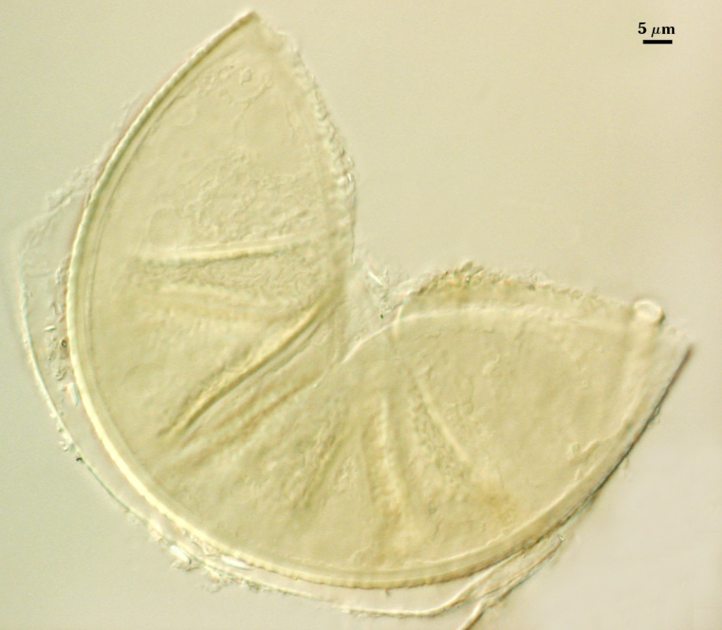
SPORE WALL: Three layers (L1, L2 and L3), all tightly adherent.
L1: Hyaline, 0.6-1.4 µm thick (mean = 1.0 µm) when intact, degrading to various degrees and sometimes sloughing completely. Often with a coating of organic debris. No obvious reaction in Melzer’s reagent.
L2: Permanent hyaline layer, 0.6-1.3 µm thick (mean = 0.9 µm), minute ridges (<0.5 µm high) on the outer surface in a finely reticulate or labyrinthian pattern. turns dark yellow (0-0-40-0) melzer's reagent.
L3: Permanent hyaline layer, 0.9-2.5 µm thick (mean = 1.8 µm) except near the subtending hypha, where it widens up to 6.9 µm.
| In PVLG | ||
|---|---|---|
 |  |  |
| In PVLG and Melzer’s reagent (1:1 v/v) | ||
|---|---|---|
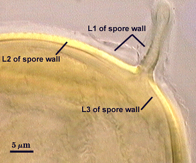 |  | 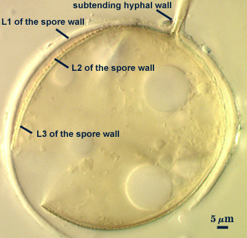 |
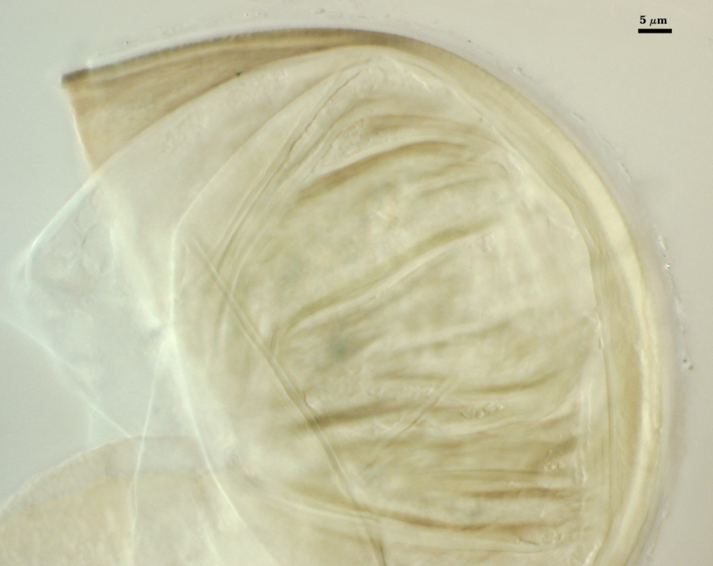
Subtending Hypha
SHAPE: Cylindrical to slightly flared (see photos above).
WIDTH: 3.8-5.6 µm (mean = 4.7 µm) at spore, narrowing to 1.6 µm distal to the spore.
HYPHAL WALL: Two hyaline layers (L1 and L2) continuous with L1 and L2 of the spore wall, but each are less than 0.5 µm thick; composite wall thickness near the spore 0.6-1.3 µm (mean = 0.9 µm). L3 of the spore wall also may be a component layer in the hyphal wall, but if so, it is not easily resolved at the light microscope level.
OCCLUSION: Closure by L3 of the spore wall (in photo at right, spore is in Melzer’s reagent).
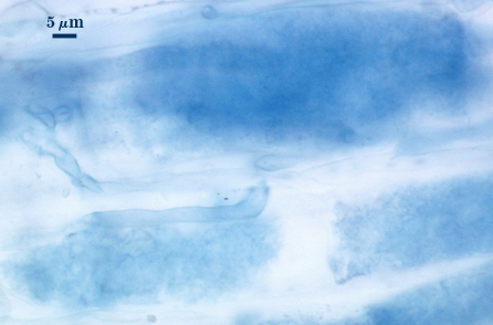
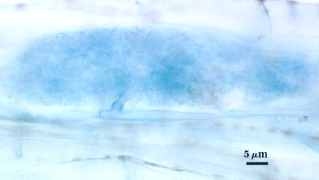
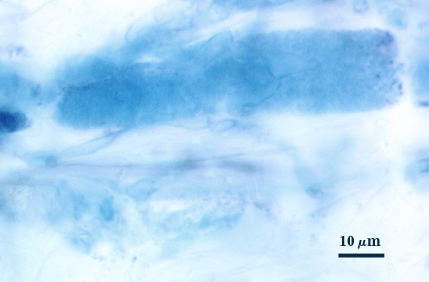
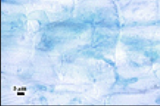
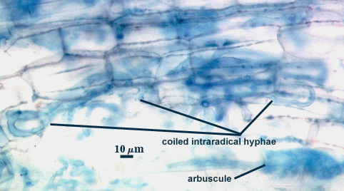
Mycorrhizae
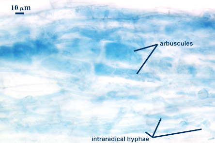
Infection units are sporadically distributed throughout colonized roots. Fungal structures most often seen are intraradical hyphae and intracellular arbuscules, but these structures stain weakly or not at all in trypan blue or other traditional acidic stains. Thus, observed patterns may not truly reflect to actual pattern of fungal distribution in roots. Amount of colonization rarely exceeds 15% of root samples of different host species (corn, red clover, leek, sudangrass), despite prolific extramatrical hyphae with spores in the rhizosphere (see right).
Appressoria evident at colonization entry points, with extensive hyphal coiling in that region. Arbuscules typically stain more faintly than hyphae, with narrow trunks (less than 4 µm wide) and extremely fine branches. Intercellular hyphae generally 2-8 µm wide, cylindrical with few knobs, irregularly branched, with some coiling. Under a stereomicroscope, faint evidence of hyphal colonization marks infection units which can then be examined more closely under a compound microscope. Of all the species producing faint mycorrhizae, P. brasilianum is the only one showing some evidence of vesicle formation. However, these subglobose, oil-filled structures also have a thicker wall than typical vesicles, suggesting they may be intraradical spores.
| Arbuscules (digitally enhanced) | ||
|---|---|---|
 |  |  |
| All mycorrhizal structures (digitally enhanced) | ||
|---|---|---|
 |  |  |
Notes
Spores are almost indistinguishable from P. occultum under a dissecting microscope; they are similar in size, shape, color (or lack of), and they also have the unusual property of floating in water. They can be separated under a stereomicroscope, but only with a very practiced eye: spores of P. brasilianum are a less reflective of light (duller looking) that those of P. occultum because of fine ornamentations that are light-scattering.
Spores of this species and P. occultum (both being members of Paraglomeraceae) are indistinguishable from those of Glomus morphologically, but rDNA and beta-tubulin genes differentiate both species and place them in a unique monophyletic clade. A region of rDNA from family members specifically is amplified using the ARCH1311 primer (TGCTAAATAGCCAGGCTGY).
The images below can be uploaded into your browser by clicking on the thumbnail or can be downloaded to your computer by clicking on the link below each image. Please do not use these images for other than personal use without expressed permission from INVAM.
High Resolution Images | |
|---|---|
|  |
 |  |
 |  |