Paraglomus occultum
(reference accession CL700) = Glomus occultum
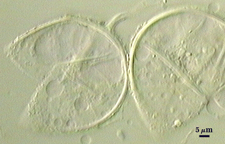
Whole Spores
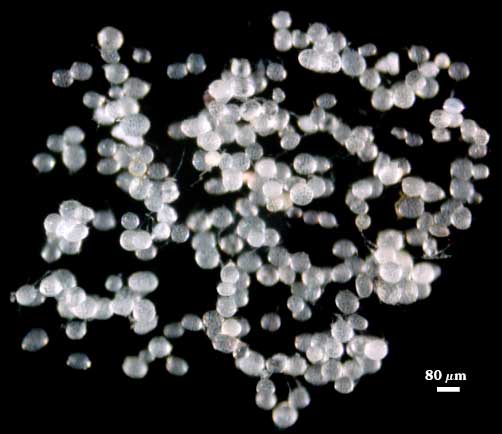
 COLOR: Hyaline (0-0-0-0) to pale cream (0-0-5-0)
SHAPE: Globose, subglobose, often slightly irregular
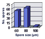
SIZE DISTRIBUTION: 60-100 µm, mean = 71.5 µm (n = 120)
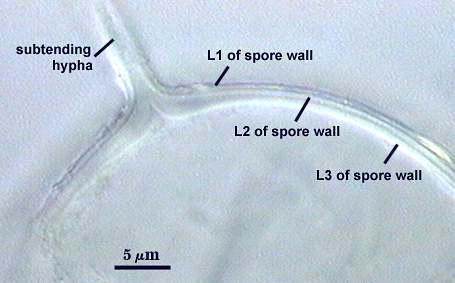
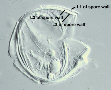
Subcellular Structure of Spores
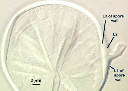
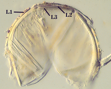
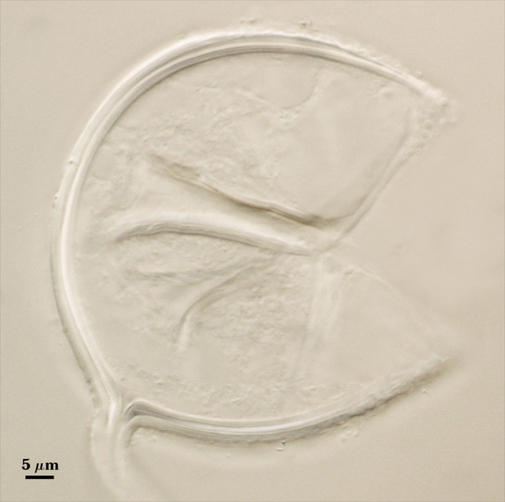
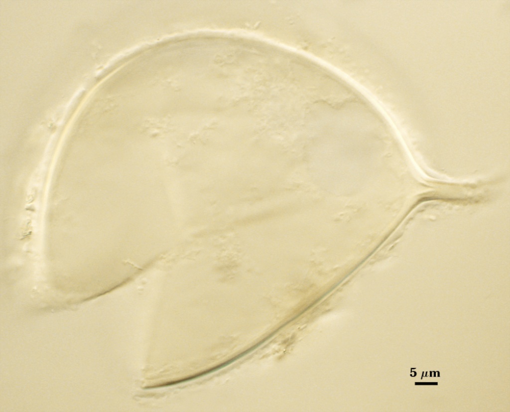
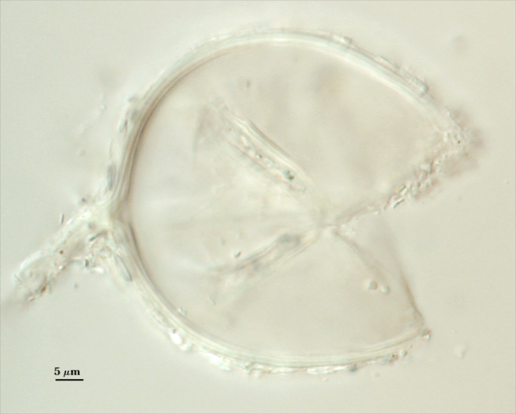
SPORE WALL: Consists of three layers (L1, L2, and L3). In the photos below, the top row is of spores mounted and broken in PVLG; the bottom row is of spores mounted and broken in 1:1 v/v PVLG and Melzer’s reagent.
L1: Generally a sloughing layer, 0.5-1.4 µm thick when intact, often separating and degrading to form a granular layer. Spores then appear to have a thin coating of organic debris on their surface and can look “dirty”. No reaction in Melzer’s reagent.
L2: A permanent layer, <0.5-1.2 µm thick (most 0.6-0.8 µm), continuing into the wall of subtending hypha without any noticeable thickening. produces a light yellow (0-0-10-0) reaction in melzer's reagent.
L3: A permanent layer, <0.5-1.2 µm thick, but often increasing in thickness to 1.6 the region of hyphal attachment, continuing into wall subtending hypha. produces a light yellow (0-0-10-0) reaction melzer's reagent.
| In PVLG | ||
|---|---|---|
 |  |  |
| In Melzer’s reagent | ||
|---|---|---|
 |  | 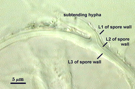 |
Subtending Hypha
SHAPE: Cylindrical to slightly flared (see photos above).
WIDTH: 3.0-5.2 µm (mean = 4.1 µm)
HYPHAL WALL: Three hyaline layers (L1, L2 and L3), continuous with layers of the spore wall, with the inner layer thickest (0.3-0.5 µm). The hyphal wall tapers to less than 0.2 µm at a distance of 5 µm or more from the spore and becomes difficult to see.
OCCLUSION: Thickening of L2 and L3 of the spore wall.
Mycorrhizae
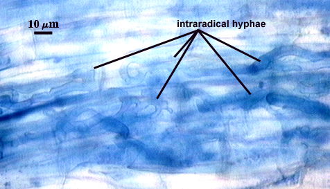
Mycorrhiza distribution appears very patchy, with spores and extramatrical hyphae often massed around root lengths with no apparent colonization. Appressoria at entry points and hyphae in the outer layers of the root cortex stain lightly in trypan blue; with some arbuscules formed near entry points also are faintly visible. Cortical colonization by hyphae, arbuscules, and vesicles are difficult to see because of poor contrast with such light staining of fungal cell walls. Arbuscules very faint, with narrow trunks (< 4 µm) and fine branching of tips. Intraradical hyphae, 2-8 µm in width, tend to coil at or near entry points; extraradical hyphae are thin, usually less than 4 µm wide. Intracellular hyphae often more coiled than intercellular hyphae. Contrast in photos below has been digitally enhanced to better visualize structures.
| Arbuscules in corn roots | |
|---|---|
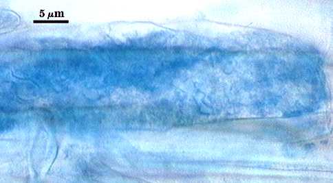 | 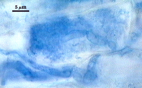 |
| All mycorrhizal structures in corn | ||
|---|---|---|
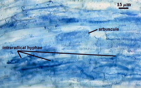 | 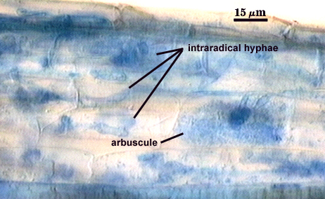 |  |
Even though mycorrhizal development is almost impossible to measure precisely because of weak staining (usually < 5% measured), it must increase with time, since sporulation increases dramatically (densities of 300 spores per cm3 of soil by some isolates). Monoclonal antibodies used to titre fungal biomass in ELISA and dot-blot assays also indicate and increase in fungal growth with time, but the details of mycorrhizal architecture and behavior still remain unknown.
Notes
Spores often float on the surface of sucrose following density gradient centrifugation and also float in water with swirling. They also tend to cling to organic debris (including spores of other arbuscular fungi).
This fungus is one of the most aggressive invaders in acid soils (particularly in forest communities) and in pot cultures (where it can be a serious contaminant if quality control measures are not taken). However, it can be found in nature almost anywhere.
Spores of this species and P. brasilianum (both being members of Paraglomaceae) are indistinguishable from those of Glomus morphologically.
The images below can be uploaded into your browser by clicking on the thumbnail or can be downloaded to your computer by clicking on the link below each image. Please do not use these images for other than personal use without expressed permission from INVAM.
High Resolution Images | |
|---|---|
|  |
 |  |