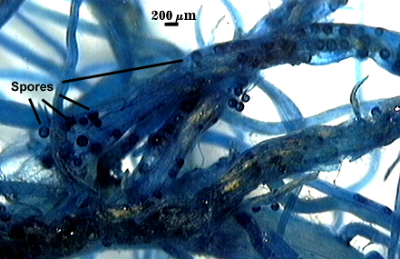
Rhizophagus clarus
(reference accession FL979A)
=Glomus clarum
=Rhizophagus manihotis
=Glomus manihotis
| Freshly extracted | Stored dry 2 years | From pigmented roots |
|---|---|---|
|
|
|
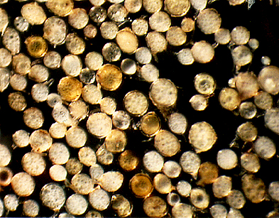
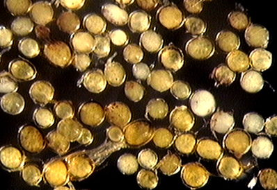
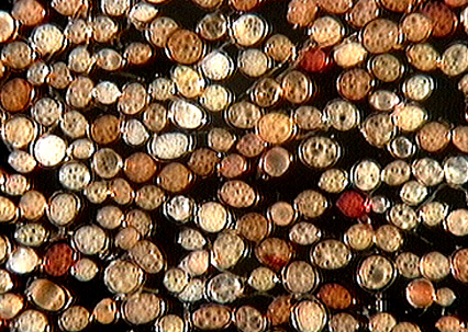
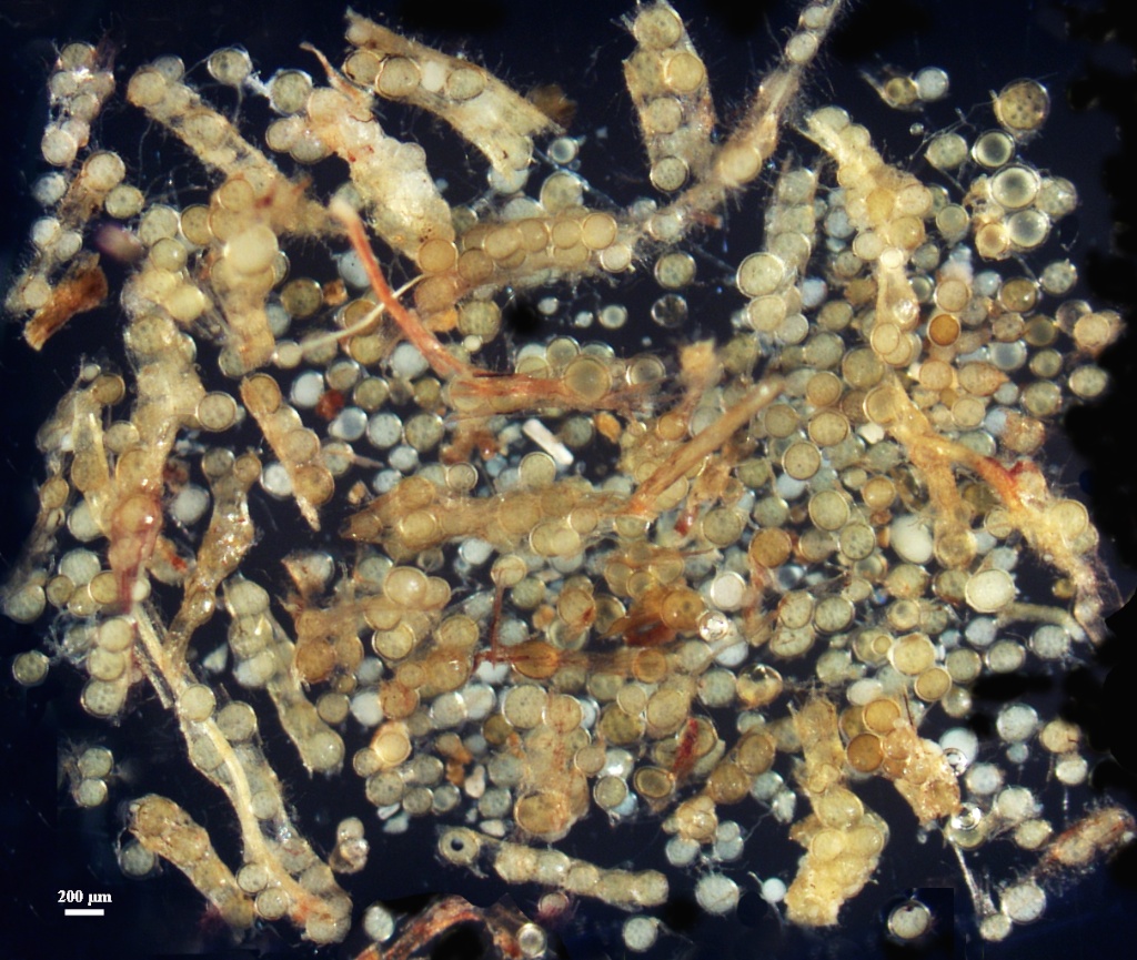
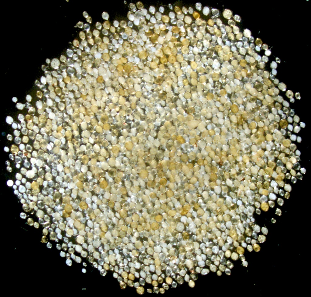
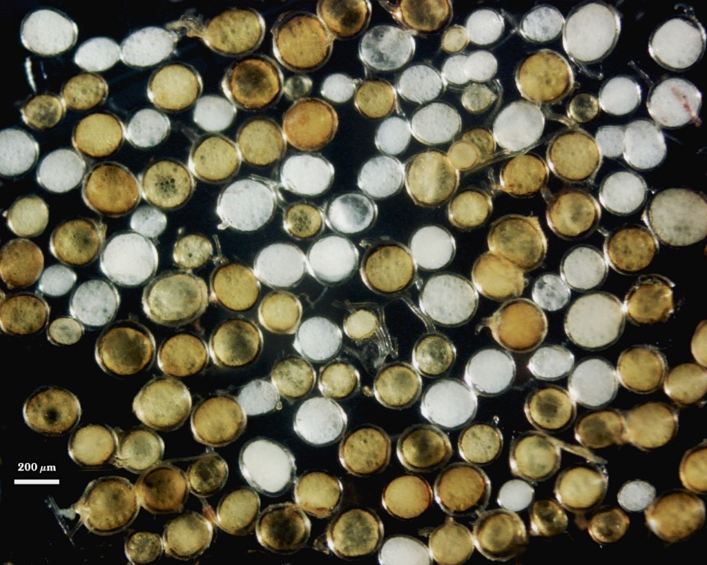
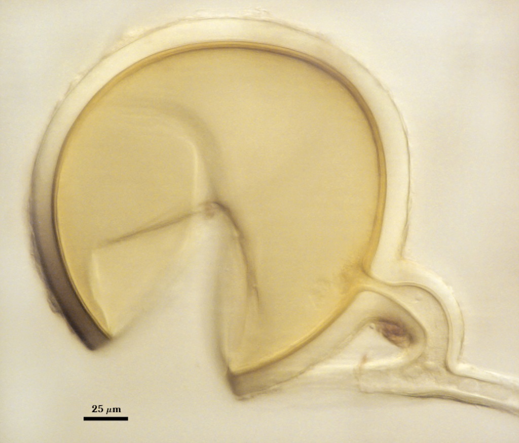
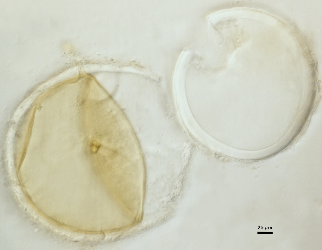
Whole Spores
 COLOR: A continuum from white to yellow-brown (0-10-60-0), with many pale yellow (0-0-20-0 to pale yellow brown (0-10-20-0).
SHAPE: Globose, subglobose, sometimes elliptical, oblong, or irregular (possibly spores formed in roots).
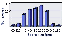
SIZE DISTRIBUTION: 100-260 µm, mean = 182 µm (n = 120).
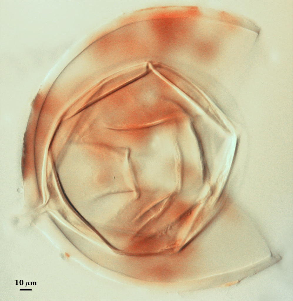
Subcellular Structure of Spores
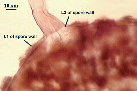
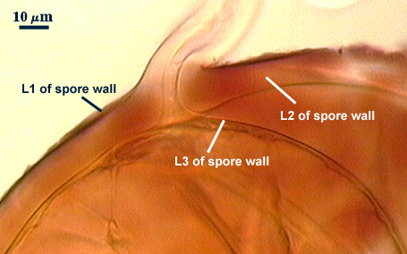
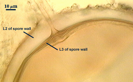
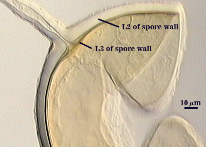
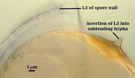
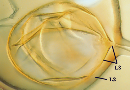
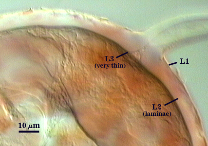
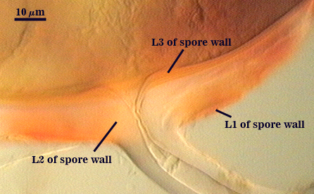
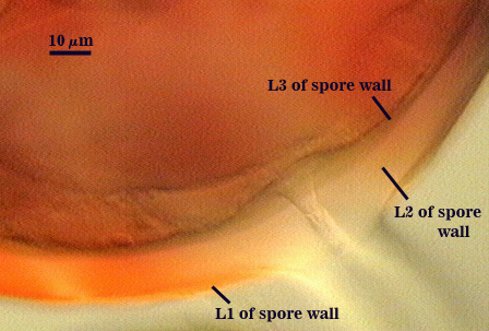
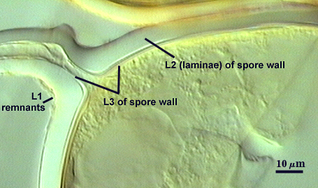
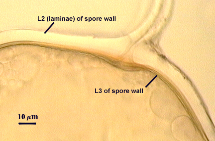
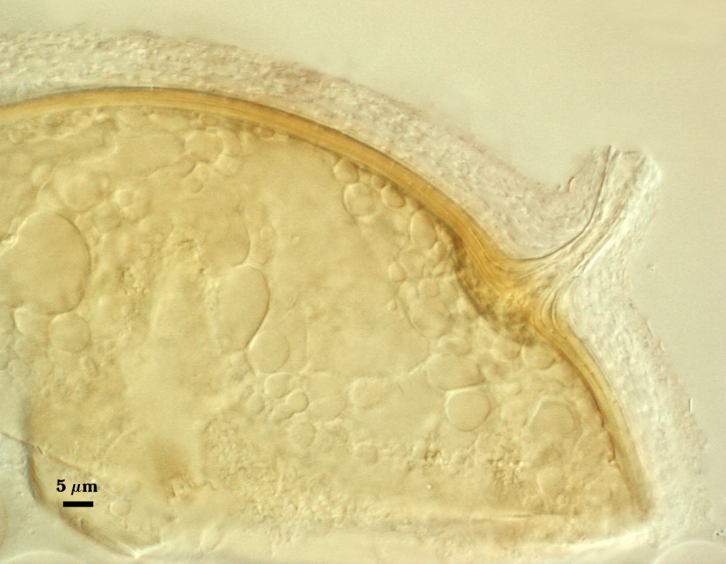
SPORE WALL: Three layers (L1, L2 and L3), all may be adherent but L3 often separates in mature spores with applied pressure. These layers form consecutively as the spore wall differentiates (left to right in sequence of photos below), with only L1 present in the spore and hyphal wall.
| Hyaline mucilagenous layer | |||
|---|---|---|---|
|
|
|
|
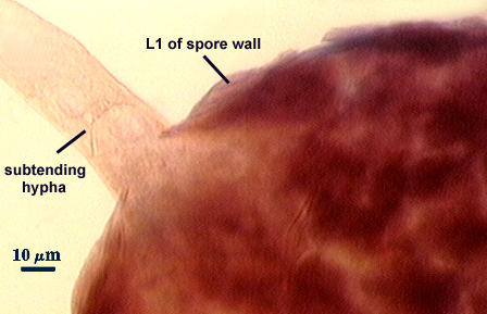
L1: A hyaline mucilagenous layer, usually degrading and sloughing so that it often is absent on mature spores; 1.0-4.7 µm thick when even distributed on spore, but often expanding in patches up to 8.5 µm thick in very young spores; staining pinkish-red (20-80-20-0) to light purple (20-80-40-0) in Melzer’s reagent. This layer almost appears laminate in juvenile spores from striations in the thicker patches.
L2: A permanent hyaline layer; 9-14.5 µm thick (mean of 11.3 µm ); consisting of sublayers that are not brittle, but rather of a granular consistency that causes cracking and fragmentation of the spore wall; spores often appear “squashed” rather than cleanly broken (see photo at right). This layer is thicker (as much as 25%) in some regions so that the inner surface appears “wavy” and sometimes appears to have shallow knobs. The surface of this layer often appears flaky in PVLG and patchy in Melzer’s reagent due to uneven degradation of the outer layer (L1).
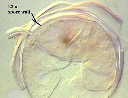
L3: Another permanent layer; usually consisting of 2-4 sublayers (or laminae); yellow (0-0-40-0) to dark yellow (0-10-60-0) in color; 1.2-3.5 µm thick (most near 2.5 µm). Sublayers usually are adherent, but they sometimes separate and thicken (up to 12 µm thick) at the juncture of the subtending hypha.
| In PVLG | ||
|---|---|---|
|
|
|
| In PVLG and Melzer's reagent (1:1 v/v) | ||
|---|---|---|
|
|
|
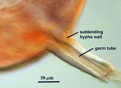
Subtending Hypha
SHAPE: Cylindrical to flared, occasionally slightly constricted or irregular.
WIDTH: 18-24 µm (mean = 22.1 µm).
WALL STRUCTURE: Three layers (L1, L2 and L3) near the spore, with L1 thinning to invisibility within 5 µm of the spore surface; composite thickness of 6.4-10.6 µm near the spore (mean of 8.7 µm), tapering to less than 3 µm with only L3 visible. L1 is mucilagenous and the only layer present in juvenile subtending hyphae, at which time the hyphal wall is < 1.0 µm thick. L2 is laminate and is continuous with L2 of the spore wall. It appears to arise de novo at the same time it is synthesized in early spore expansion; 4.8-11.2 µm thick near the spore at maturity. L3 is visible as a very thin layer (usually < 0.8 µm thick, occasionally up to 1.6 µm) and pale yellow (0-0-10-0). In some white spores, this layer cannot be seen.
OCCLUSION: Innermost sublayer of L3 of the spore wall bridges the pore, although often it is so thin that the pore appears to be open (see photos above). In some spores, the spore wall thickens and almost occludes the pore; a plug is formed with rare frequency.
| Clarus Spore Wall | |
|---|---|
|
|
Germination
A germ tube emerges from the lumen of the subtending hypha, and originates either at the septum or at a break in branch hyphae. It appears to arise from the innermost sublayer of L3 in both cases.
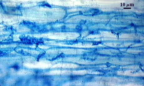
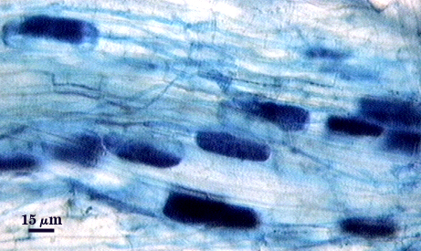
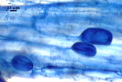
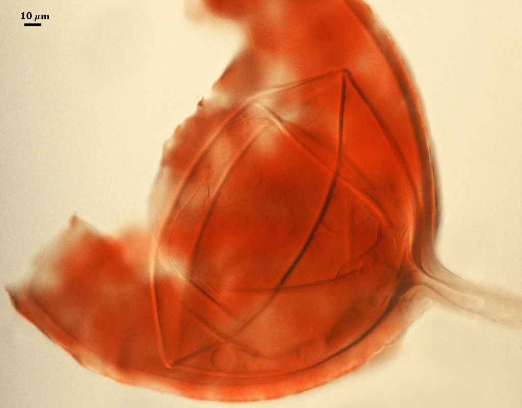
Mycorrhizae
| Arbuscules in corn root cells | |
|---|---|
|
|
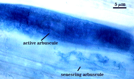
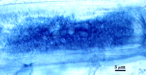
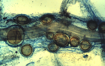
Elliptical to oblong vesicles often are produced near point of entry into a root, after which they quickly form a thickening outer wall that differentiates into larger spores. Arbuscule production seems to peak early in mycorrhizal colonization (photos below are at 5 weeks after corn emergence), with arbuscules forming in isolated cells so that the distribution appears dispersed and patchy. Intraradical hyphae range from 2-5 µm in diameter, increasing in thickness if a terminal vesicle (spore) forms. Hyphae often grow for some distance in parallel to each other, connected by distinct “H” and “h” branching patterns. All structures stain darkly in Melzer’s reagent except for senescing arbuscules (see below). Intraradical spores often congregate in clusters and break through the root epidermis because of their large size.
| All mycorrhizal structures in corn roots | ||||
|---|---|---|---|---|
|
|
|
|
|
Notes
The range in spore size and color is greater than that found for most glomoid species, with the latter attributed to the thickness of the second laminate layer of the spore wall (L3), which is very thin in white spores and much thicker in yellow to yellow-brown spores. Some isolates in INVAM are consistently white, but the spores are at the small end of the size distribution, others are highly variable in color (changing with pot culture generation), and still others are mostly yellow to yellow-brown. This continuum eliminates any morphological distinction between R. clarus and Rhizophagus manihotis.
The images below can be uploaded into your browser by clicking on the thumbnail or can be downloaded to your computer by clicking on the link below each image. Please do not use these images for other than personal use without expressed permission from INVAM.
High Resolution Images | |
|---|---|
 |  |
 |  |
 |  |
 |  |