Acaulospora colombiana
(reference accession CL356)
= Entrophospora colombiana
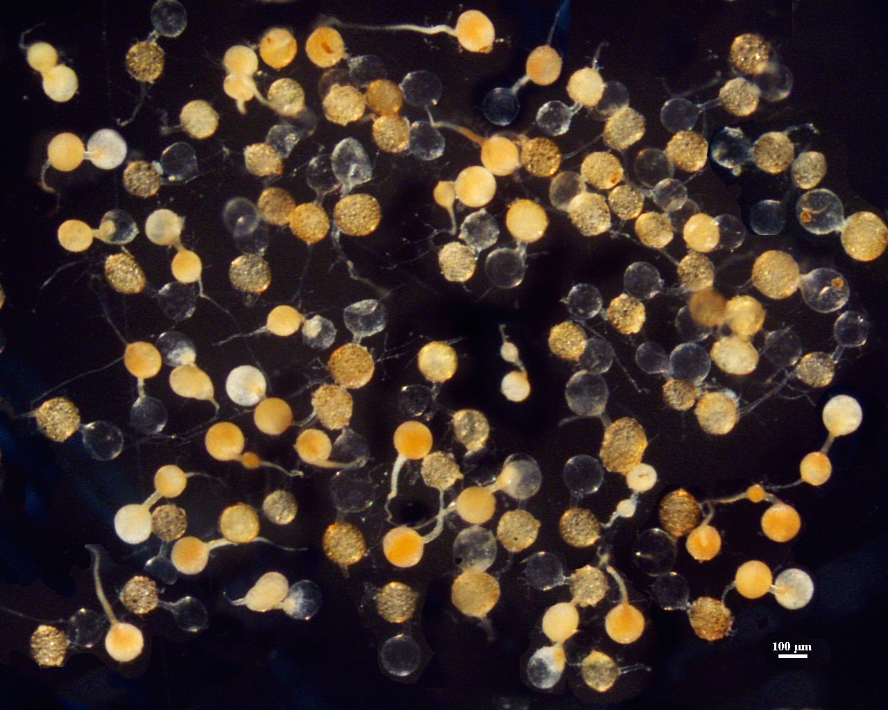
Whole Spores
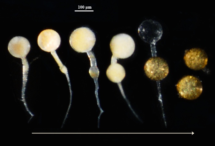
Below photo here shows appearance of spores from the time they first begin to form (2) on a sporiferous saccule (1) until they mature and become sessile (3-5).
| Spore Growth | |
|---|---|
 | 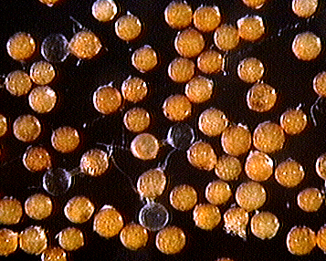 |
 COLOR: Orange-brown (0-20-60-0) to dark orange-brown (0-10-60-0), many between these two extremes (0-40-100-0). The contents of immature spores are dense, so spores appear much more opaque than at maturity.
SHAPE: Globose to subglobose.
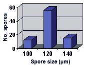
SIZE DISTRIBUTION: 100-140 µm, mean = 121 µm (n = 83).
Subcellular Structure of Spores
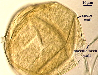
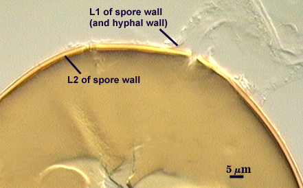
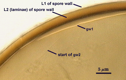
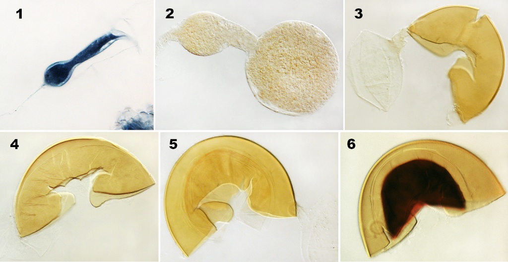
SPORE WALL: Two layers (L1 and L2), the outer layer continuous with the wall of the neck of the parent sporiferous saccule and the inner layer initiates origination of the spore. Below (left to right) is the sequence in differentiation of all spore subcellular structures.
| Differentiation Sequence | ||
|---|---|---|
 |  |  |
| Differentiation of all spore subcellular structures | |||
|---|---|---|---|
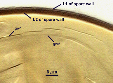 |  |  | 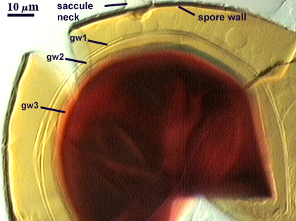 |
L1: A hyaline layer that is continuous with the wall of the saccule neck; 0.5-0.8 µm thick; usually present on spores with saccules attached and sloughed when saccules have detached.
L2: A layer consisting of yellow-brown (0-30-100-0) to darker yellow-brown (0-30-100-10) sublayers (or laminae) that originates from L1 as the spore expands. Thickness ranges from 2.4-4.4 µm thick (mean of 3.3 µm). At maturity, the pore between spore and saccule neck is closed by continuous sublayers of this layer to form an “endospore”.
L3: A single layer, 0.5-0.6 µm thick, probably concolorous (difficult to determine due to thinness). Its position is highly variable depending on how much a spore is broken, which has caused many years of confusion in interpretation of whether it is part of the spore wall or not. It separates to varying degrees from the spore wall, but rarely is detectable when adherent. When separation is uniform around the spore wall, this layer can be misinterpreted as a flexible inner wall. However, it is slightly more rigidity than flexible inner walls because it can break along with the spore wall (thus its previous definition as “semi-rigid”), and also can sometimes have sublayers of varying numbers.
| Spores mounted in PVLG | |
|---|---|
|
|
| Spores in PVLG & Melzer’s reagent | |
|---|---|
|
|
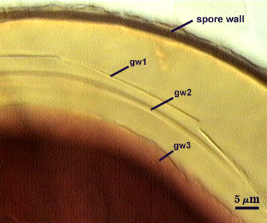
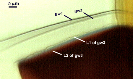
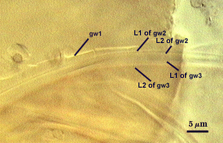
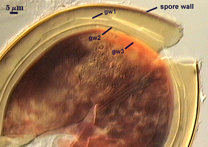
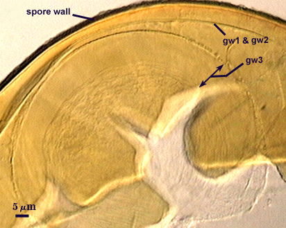
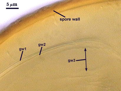
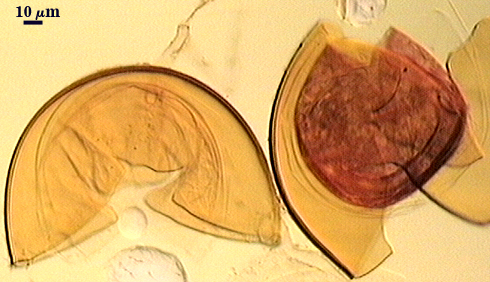
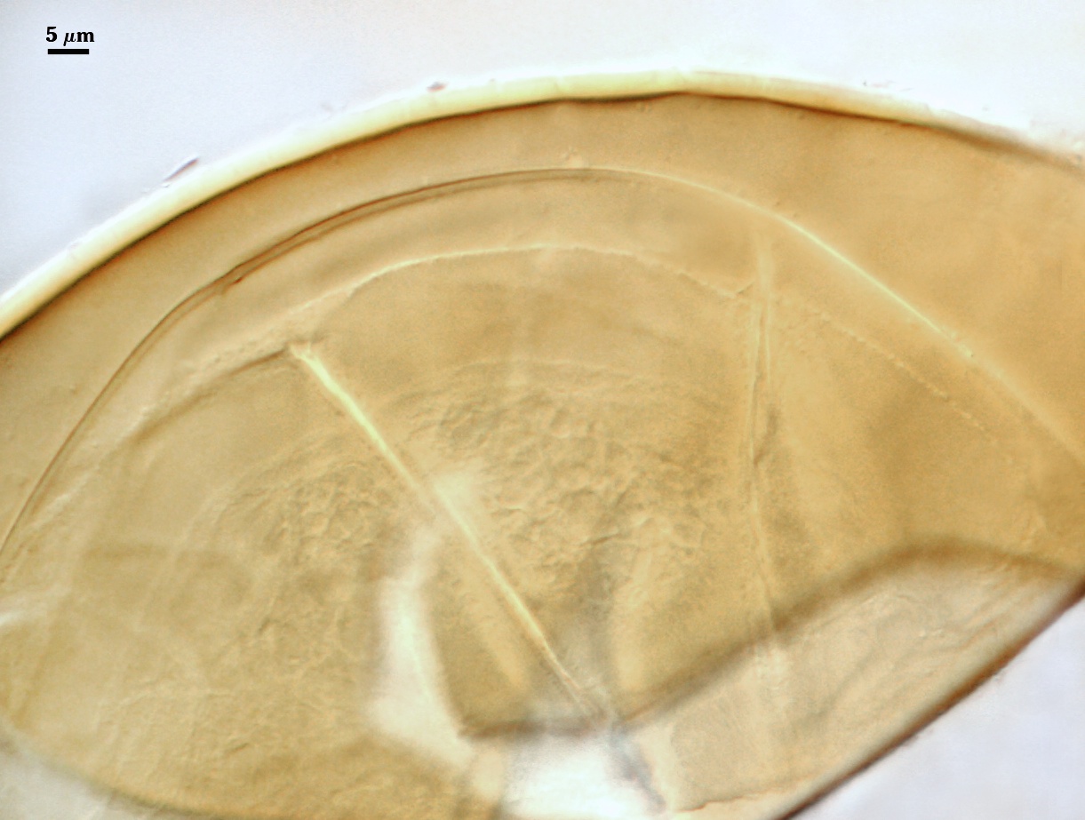
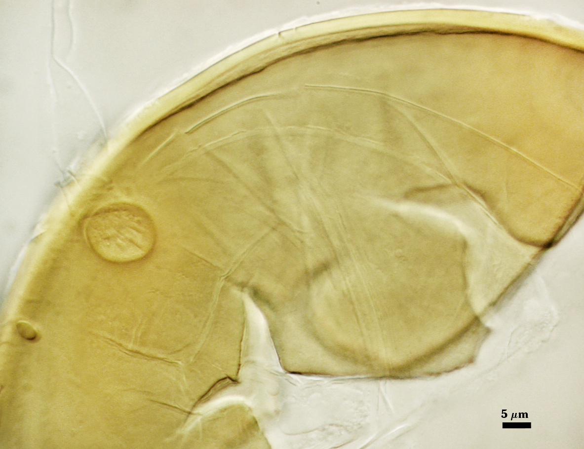
GERMINAL WALLS: Two flexible hyaline inner walls (gw1 and gw2) can be seen in all spores IF they separate when a spore is broken and they do not interfold and overlap to make them indistinguishable (and often appearing as many more than are actually present). Both gw1 and gw2 tend to separate much more in PVLG with Melzer’s reagent than in PVLG alone (see photos above). These walls form separately and sequentially only after the spore wall has completely differentiated.
GW1: Usually appears as a single layer, 0.6-1.4 µm thick, but it appears to consist of two very thin layers that almost always are adherent.
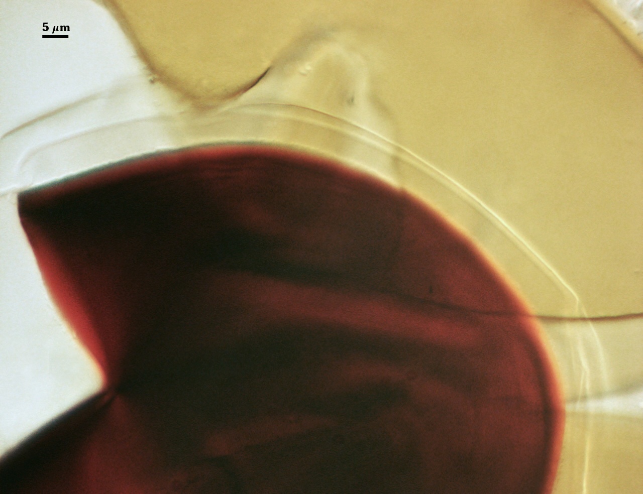
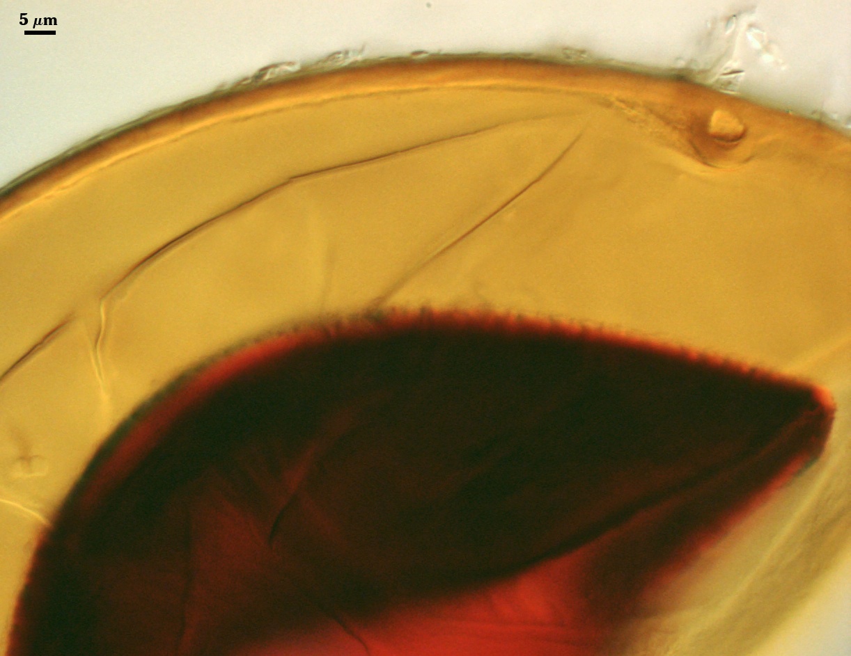
GW2: flexible wall consisting of two tightly adherent layers. L1 is hyaline, 0.5-0.8 µm thick, with granular excrescence (or “beads”) that tends to become dislodged and float away with applied pressure. These “beads” are stabilized after preservation in formalin, but otherwise may be absent on mounted spores within a few months of storage. L2 is hyaline and plastic enough that it has been termed “amorphous”. It can range in thickness from 9-20 µm in PVLG-based mountants, depending on amount of pressure applied to it while breaking the spore; staining red-purple (20-80-20-0) to dark red-purple (40-80-60-0) in Melzer’s reagent. This layer is nonreactive in youth and gradually becomes more reactive as differentiation is completed (see photo below).
Cicatrix
A circular or ovoid scar that remains from the connection between spore and saccule neck; 17-31 µm (mean = 21.7 µm) for the cicatrix proximal to the saccule, and 6-15 µm (mean = 10.8 µm) for the cicatrix distal to the saccule.
Sporiferous Saccule
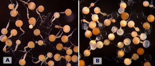
COLOR: Salmon colored in youth (A in photo at right), gradually becoming white and then hyaline as the contents are hydrolyzed or disintegrate (B in photo at right).
SHAPE: Subglobose to slightly oblong.
SIZE: 100-110 µm, mean = 102.4 µm.
SACCULE WALL: One hyaline layer, 1.0-1.4 µm; surface smooth.
DISTANCE FROM SACCULE TO SPORE: 20-70 µm.
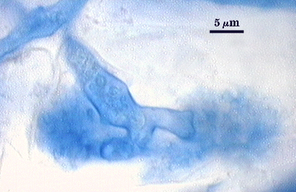
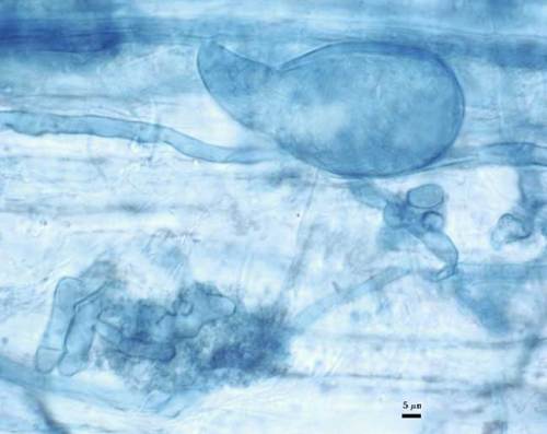
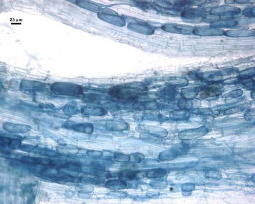
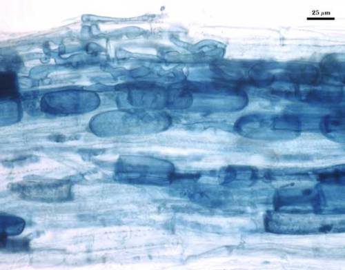
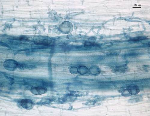
Mycorrhizae
Arbuscules generally stain lightly in trypan blue, making observation of fine architecture difficult. Intraradical hyphae and vesicles stain with much more varying intensity, from almost invisible to quite dark. Infection units appear to be patchily distributed in red clover, sudangrass, and corn, with oblong to irregular vesicles often forming in localized clusters.
| Arbuscules in cortical cells of corn | ||
|---|---|---|
|
|
|
| Mycorrhizae in corn (with distribution of infection units and vesicles). | ||
|---|---|---|
|
|
|
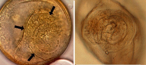
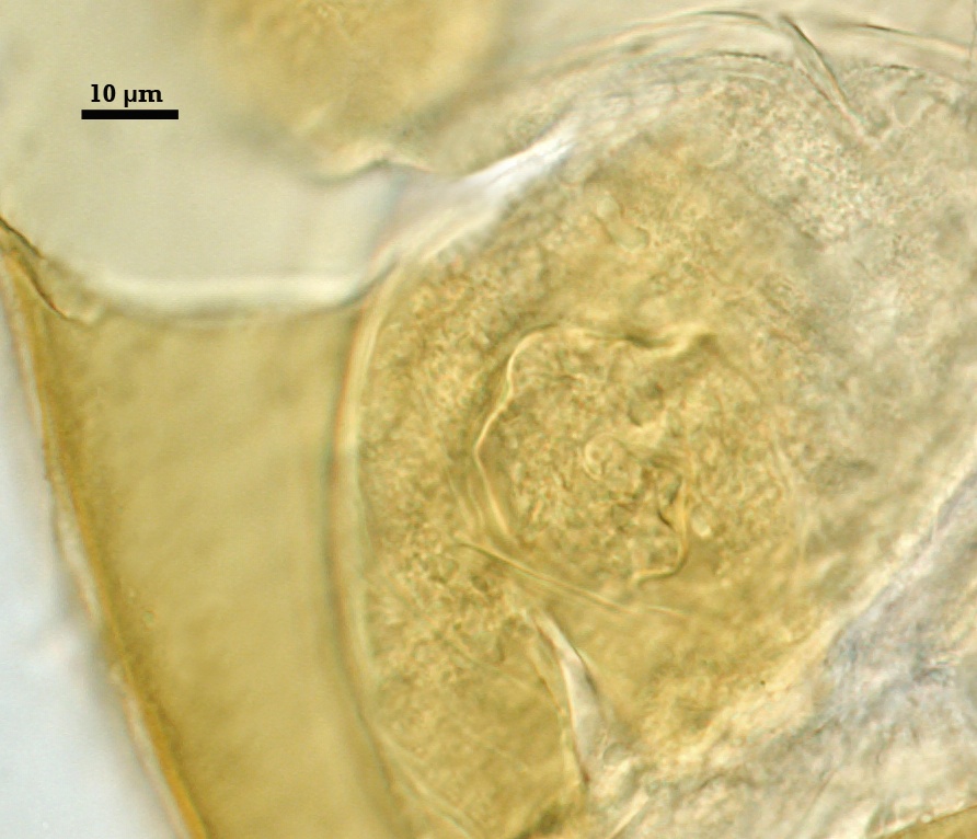
Germination
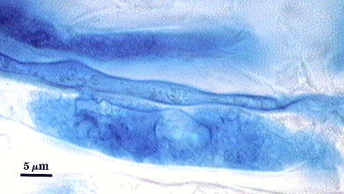
Spain (1992) reports orbs in E. colombiana spores to be slightly ovoid with an inward spiral configuration and simpler than the ones found in Acaulospora species. This orb forms on gw2, from which germ tubes form and penetrate through the spore wall. They are difficult to see except in older spores where contents have cleared with fusion of lipid globules in the spore lumen, mostly because each is wide enough to span most of the diameter of a spore. The photo at right was provided courtesy of Joyce Spain.
Notes
Spores stored in dry soil often will have an indented surface; this will disappear after 24 hr incubation in water.
Median and mean distance from spore to saccule are 30 and 36 µm, respectively. A few spores are atypically dark red-brown (0-40-100-0). The smaller of the two cicatrices has a collar 1-1.3 µm high. The large cicatrix is recognized by a small lip as a remnant from where the saccule neck branched during spore formation.
The differentiation sequence in spores is exactly that of A. mellea, with the same subcellular structure and organization. When only sessile spores are extracted from field or pot culture soils, these two species can be distinguished by the number of scars (cicatrices) on the spore wall surface.
High Resolution Images | |||
|---|---|---|---|
 |  | ||
 |  | ||
 |  | ||
 |  | ||
Reference
- Spain, J. L. 1992. Patency of shields in water-mounted spores of four species in Acaulosporaceae (Glomales). Mycotaxon 43:331-339.