Funneliformis coronatum
(reference accession AU202)
Whole Spores
| Coronatus Spheres | |
|---|---|
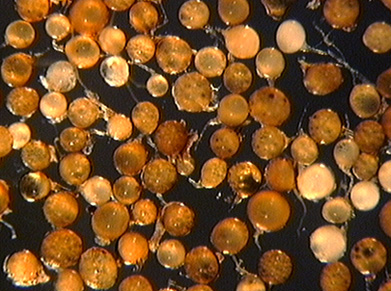 | 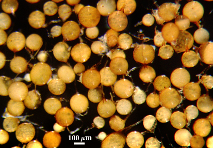 |
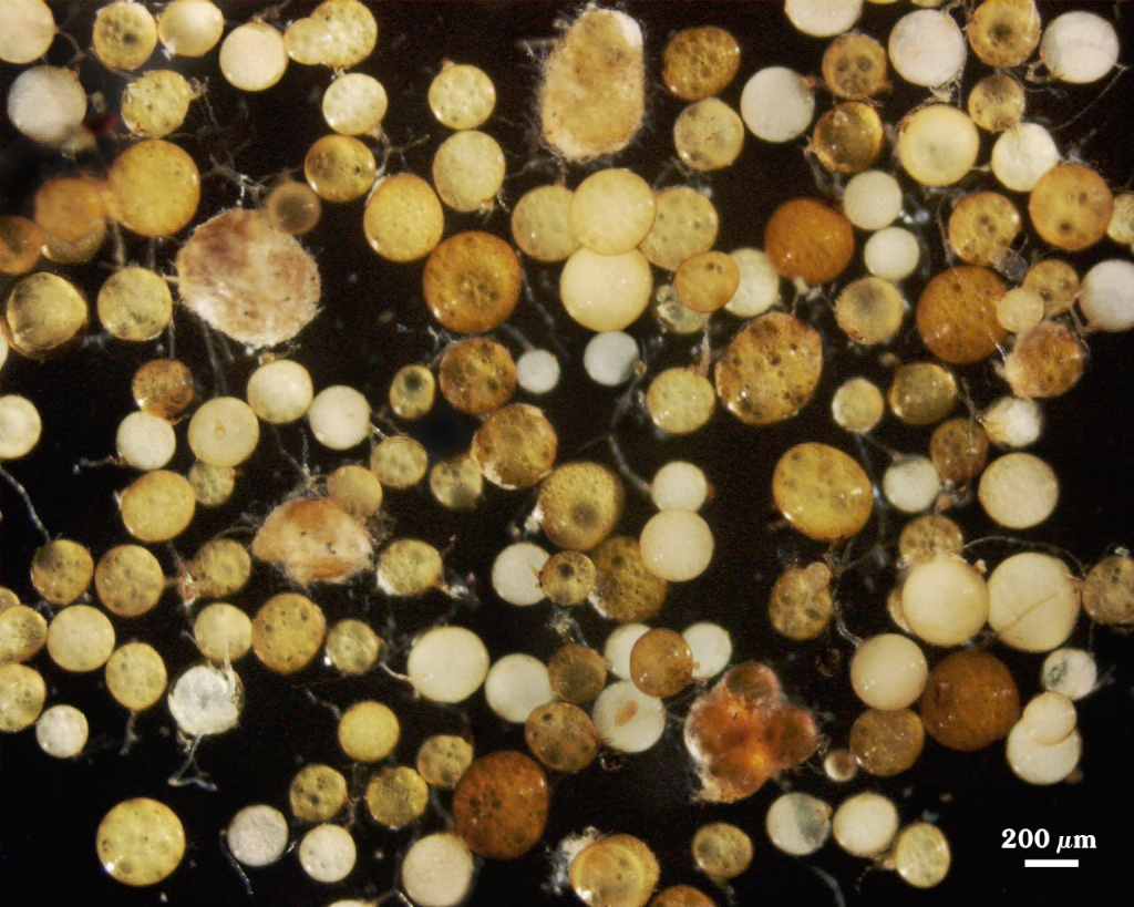
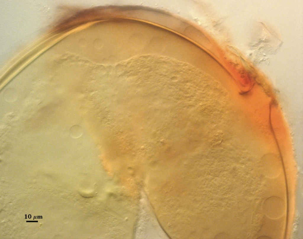
 COLOR: Pale orange-brown (0-20-60-0) to dark orange-brown (0-60-100-10), with most orange brown (0-40-80-0).
SHAPE: Globose, subglobose, some irregular.
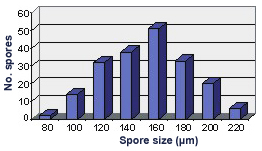
SIZE DISTRIBUTION: 80-220 µm, mean = 154 µm (n = 196).
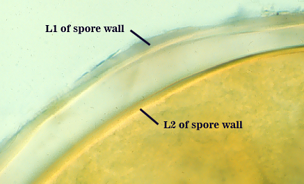
Subcellular Structure of Spores
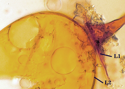
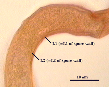
SPORE WALL: Two layers (L1 and L2), the outer one often absent in mature spores.
| In PVLG | |
|---|---|
 |  |
| In PVLG and Melzer's reagent (1:1 v/v) | |
|---|---|
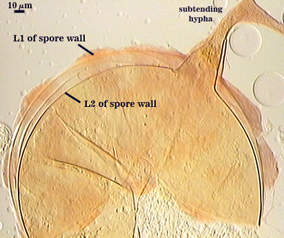 | 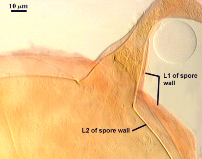 |
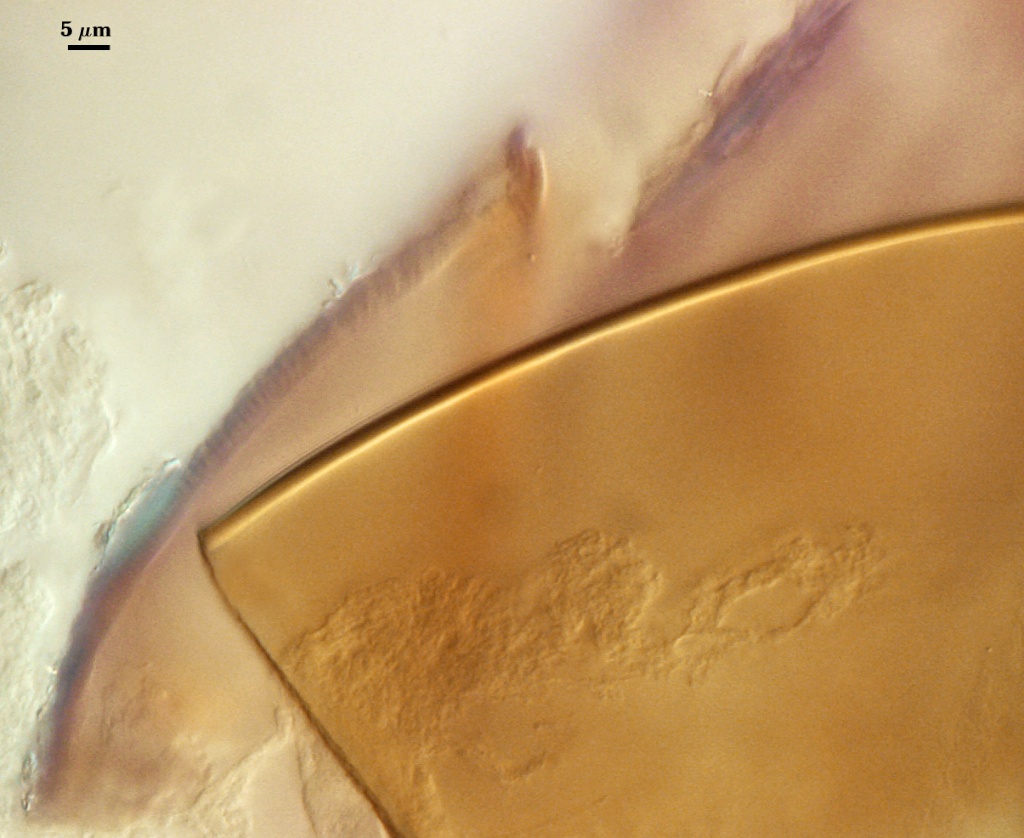
L1: Outer layer, hyaline, the surface staining pinkish-red in Melzer’s reagent (0-20-20-0 to 0-60-20-0); sloughing in mature spores, appearing granular in advanced stages of degradation; 2.5-3.5 µm thick when intact on juvenile spores.
L2: A laminate layer consisting of light orange-brown (0-20-80-0), 3.2-6.5 µm thick (mean of 5.9 µm); with smooth surface after outer layer has sloughed.
Subtending Hypha | |
|---|---|
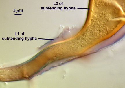 |  |
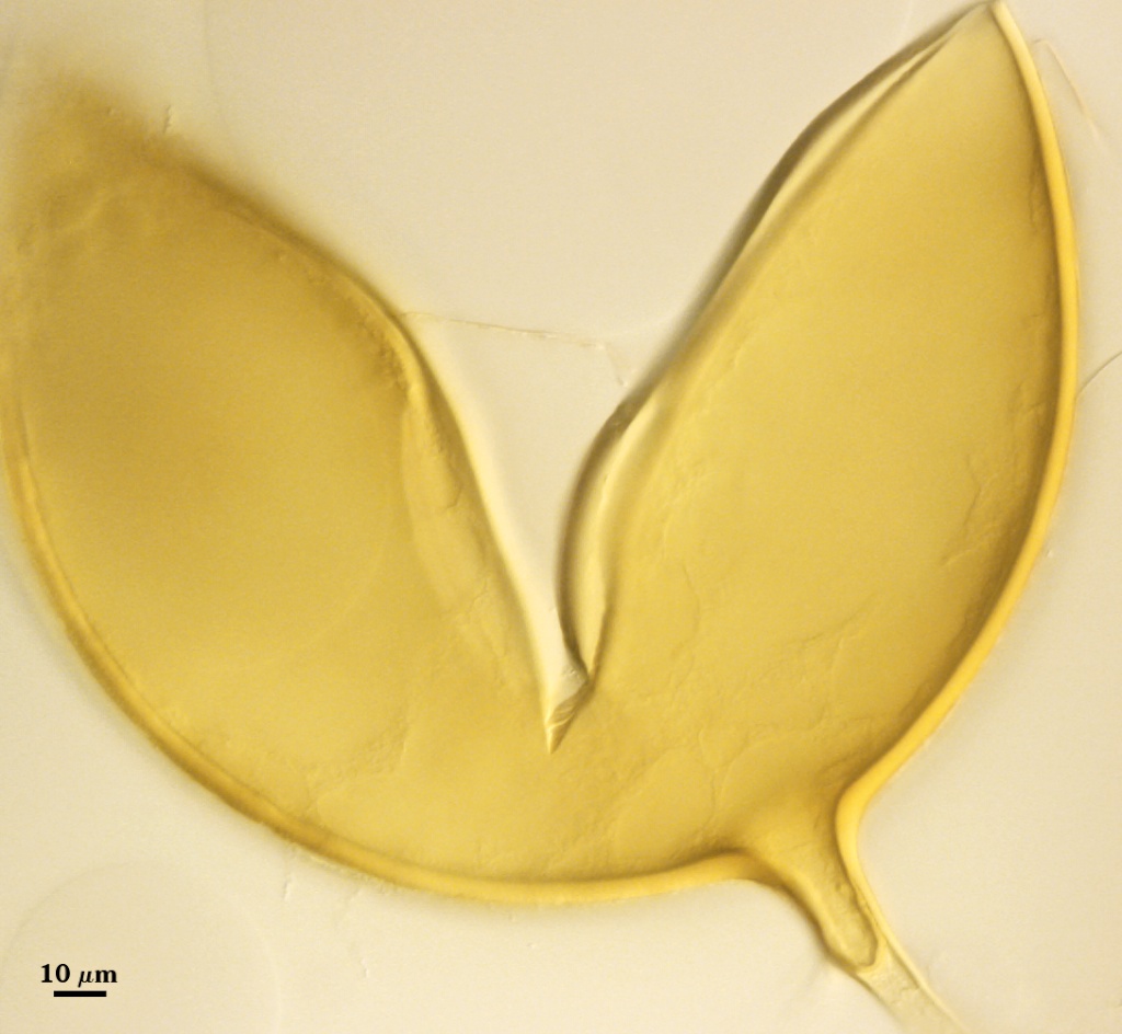
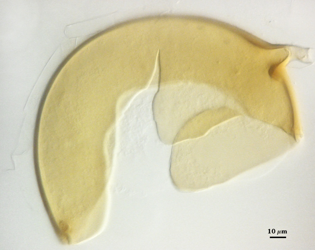
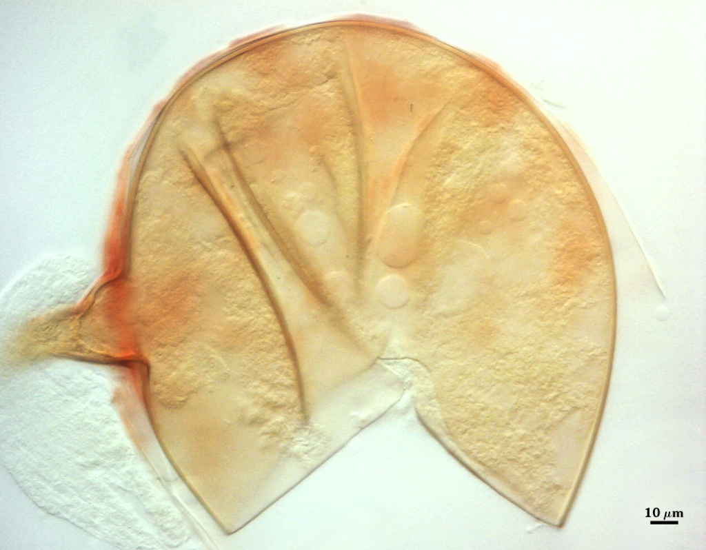
SHAPE: Markedly funnel-shaped in over 90% of spores in a population.
WIDTH: 30-42 µm (mean = 34.6 µm).
WALL STRUCTURE: Two layers (L1 and L2) continuous with the two layers of the spore wall, 3.2-7.5 µm thick (see photo at right). L1 is the outer layer, thins to 1.2-1.6 µm within 50 µm of the spore. Rarely present on the hypha of mature spores. L2 is concolorous with the laminate layer of the spore wall, sublayers adherent.
OCCLUSION: Many spores appear to have an open pore, but a thin curved septum is formed 40-60 m from attachment point; often hard to see (< 1 µm thick) because of distance from spore. In some spores, a thicker recurved septum forms nearer the point of attachment (15-20 µm).
Germination
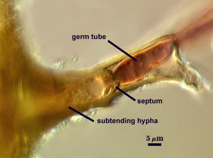
A germ tube emerges from the lumen of the subtending hypha. It appears to originate from the innermost sublayer of the subtending hypha that curves to form the septum, regardless of where that septum forms.
Mycorrhizae
Not examined yet.
Notes
The description of this species (Giovannetti et al., 1991) mentions formation of sporocarps by this species, but none were produced in pot cultures of this isolate. Moreover, spores were considerably smaller than the range reported in the protologue (173-336 µm).
The difference between this species and F. mosseae are different than those mentioned in the protologue. The outer layer of the spore wall is described as having “radiating columns” (hence the name coronata from “corona”), but this phenotype is more a product of environment and therefore is an artifact of the mounting medium rather than a heritable trait (the only kind that is important taxonomically). We do not see this trait when spores are mounted in our standardized medium, PVLG or PVLG and Melzer’s reagent. Other differences not mentioned or not emphasized by Giovannetti et al. (1991) are: (1) consistently darker brown spores, (2) a more marked funnel-shaped subtending hyphae in most, if not all, spores in a population, and (3) the absence of a slate-like layer sandwiched between L1 and L2 of the spore wall (which causes the surface of F. mosseae spores to have numerous small depressions or pits).
The images below can be uploaded into your browser by clicking on the thumbnail or can be downloaded to your computer by clicking on the link below each image. Please do not use these images for other than personal use without expressed permission from INVAM.
High Resolution Images | |
|---|---|
 |  |
 |  |
 |  |
Links to Gene Sequences in Genbank
Reference
- Giovannetti, M., L. Avio, and L. Salutini. 1991. Morphological, cytochemical, and ontogenetic characteristics of a new species of vesicular-arbuscular mycorrhizal fungus. Can. J. Bot. 69:161-167.