Gigaspora decipiens
(reference accession AU102)
| Gigaspora decipiens | |
|---|---|
|
|
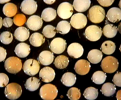
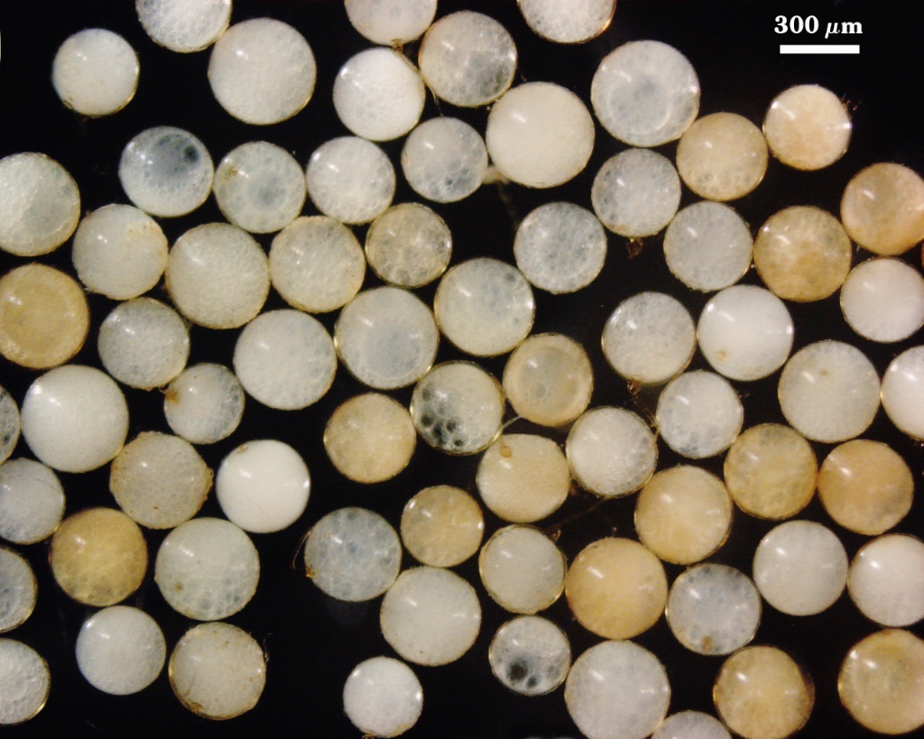
Whole Spores
 COLOR: White to cream (0-5-30-0), becoming yellow-brown (0-10-80-10) with age or prolonged storage.
SHAPE: Globose to subglobose.
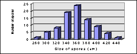
SIZE DISTRIBUTION: 280-440 µm, mean = 358 µm (n = 85).
Subcellular Structure of Spores
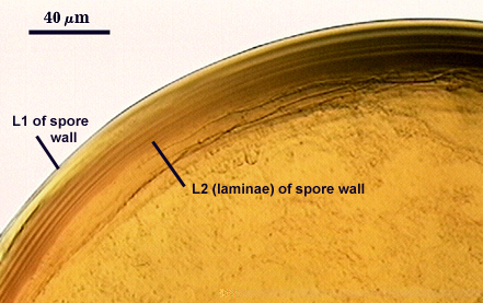
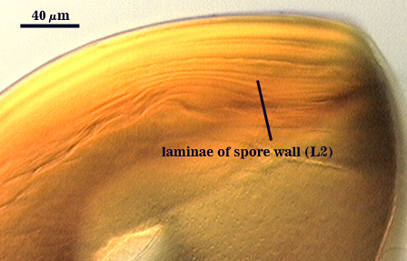
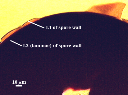
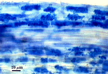
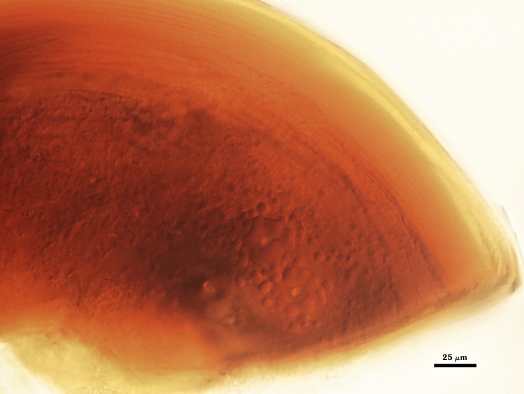
SPORE WALL: Consisting of three layers (L1, L2, and L3). The first two are adherent and of equal thickness in juvenile spores, with L2 thickening as the spore wall is differentiated; L3 differentiates as a prelude to germ tube formation.
L1: An outer permanent rigid layer, smooth, adherent to sublayers of L2, 2.5-3.2 µm thick, often hard to see in relation to L2.
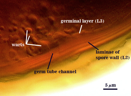
L2: A layer consisting of hyaline sublayers (or laminae) that increase in number with thickness, are rigid, exhibit some plasticity when broken, pale yellow (0-0-10-0) to yellow (0-0-40-0) in newly formed spores, turning a darker brownish yellow (0-10-60-0) with age and storage; 15-60 m thick (mean of 34.8 µm). Variation in thickness is function of number of sublayers (5-20, each 2-3 m thick) and the degree of separation between them when pressure is applied. Sublayers stain dark red-purple (almost black) in Melzer’s reagent (60-80-70-10). In younger spores, sublayers merge and resemble “waves” without sharp transitions from ridge to trough. The degree of separation between sublayers gives the impression of separate layers, but they all originate as part of the same structure and phenotype.
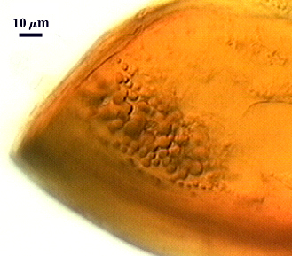
L3: A “germinal” layer that is concolorous and adherent with the laminate layer. This layer is distinct only at the ultrastructural level, where it appears electron dense. Numerous “warts” or “papillae” form on the inner surface of this layer, and they are especially concentrated in regions where germ tubes form (usually in close proximity to the suspensor cell); warts 1.2-5 µm high in germinating spores and 2.5-3 µm wide.
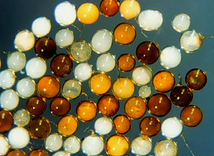
| Spores in PVLG | |
|---|---|
|
|
| Spore in 1:1 v/v PVLG and Melzer’s reagent | |
|---|---|
|
|
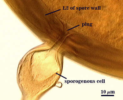
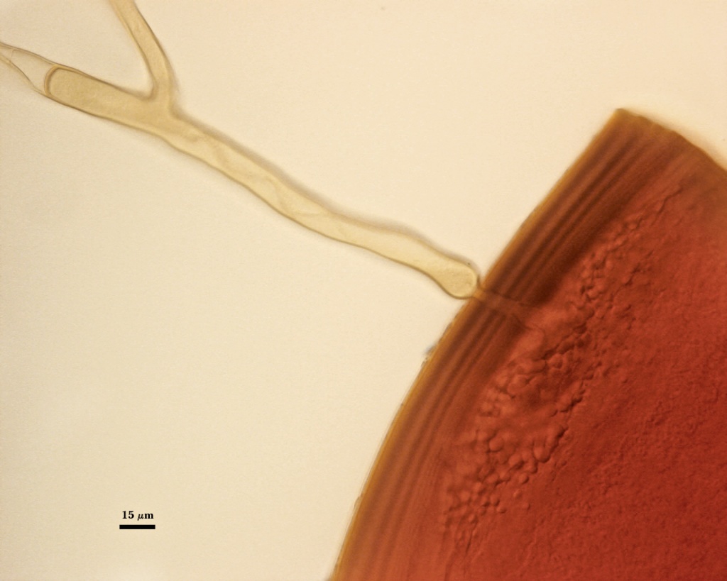
Subtending Hypha
WIDTH OF SPOROGENOUS CELL: 51-63 µm (mean = 57 µm).
SPOROGENOUS CELL WALL: 2 hyaline layers (L1 and L2) probably are present (continuous with the first two layers of the spore wall), but only L2 is readily discernible at the level of the compound microscope.
L2: Brownish yellow (0-10-80-0), 5-6.8 µm thick near the spore and then thinning to 1.2-2.0 µm beyond the sporogenous cell.
OCCLUSION: Closure by a plug concolorous with the laminate layer of the spore wall (see photo).
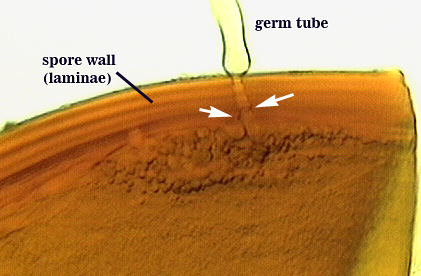
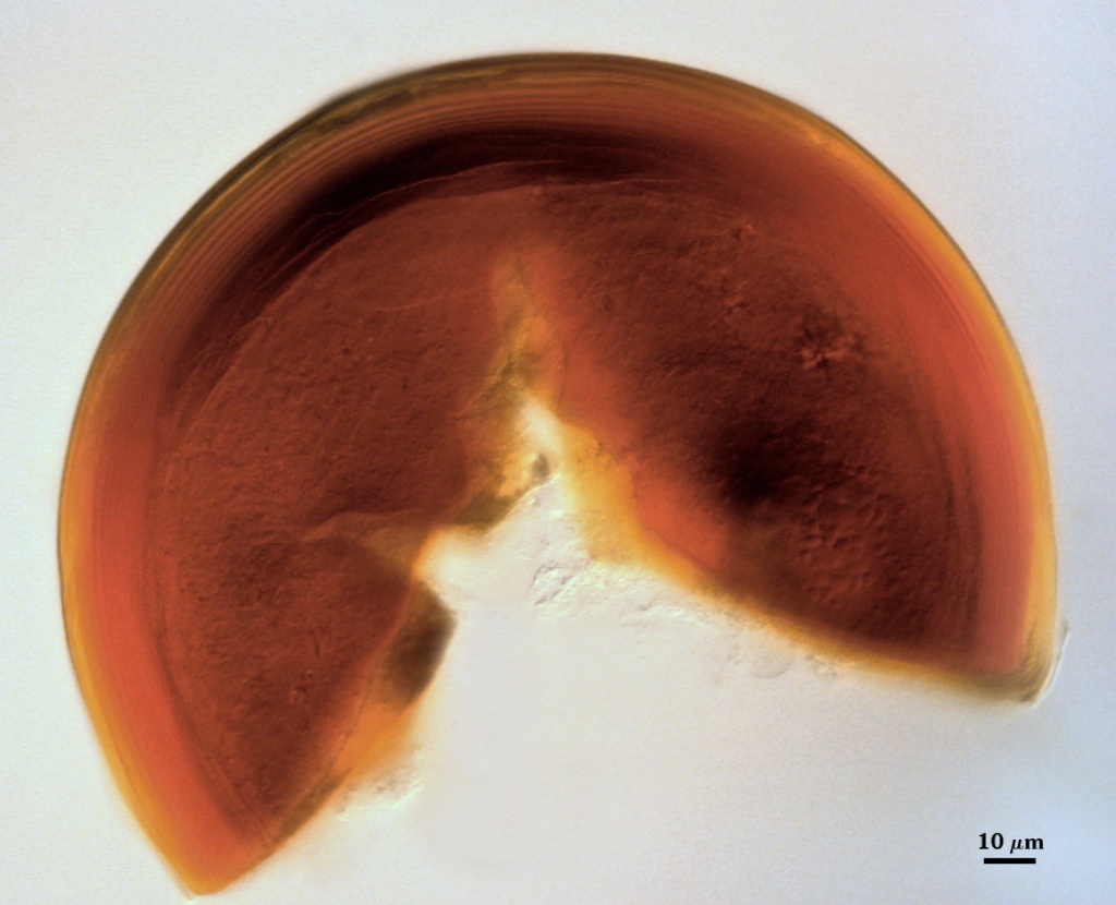
Germination
Germ tubes forms in vicinity of warty protruberances that make up the innermost layer (L3) of the spore wall (right photo below). The germ tube hole that passes through all layers of the spore wall is 6-9 µm wide (see white arrows in the left photo), with the germ tube expanding immediately after emergence from the spore wall (14-16 µm wide).
| Spores in PVLG | ||
|---|---|---|
|
|
|
The spore wall originally was pale yellow-brown) but deepened to orange-brown as a result of storage in this mountant.
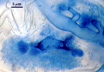
Mycorrhizae
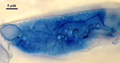
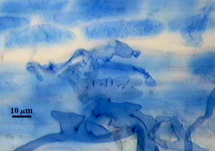
Intraradical arbuscules and hyphae consistently stain darkly in roots treated with trypan blue. Arbuscules with many fine tips from a swollen and often coiled trunk hypha. Extraradical and intraradical hyphae often with knobs or projections, the thicker hyphae ranging from 4-8 µm in width (most 4-6 µm); intraradical hyphae usually densely coiled near entry points.
| Arbuscules in cortical cells of corn | |
|---|---|
|
|
| Views of trypan blue-stained corn roots under a dissecting scope | |
|---|---|
|
|
| External hyphae, entry points, and arbuscules in corn mycorrhizae | |
|---|---|
|
|
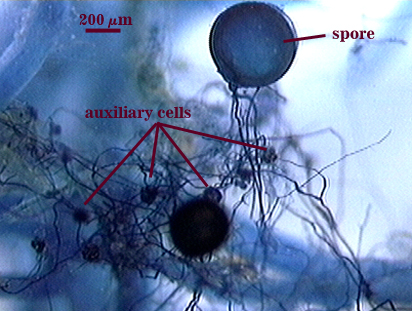
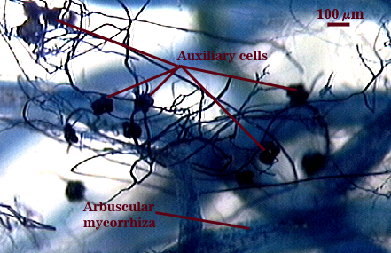
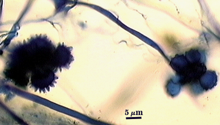
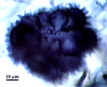
Auxiliary Cells
Soil-borne auxiliary cells form in aggregates of 1-10 cells borne together on a tightly coiled hyaline hypha (branching from a parent extramatrical hypha). Each cell is thin-walled (< 1 µm thick), pale cream (0-0-10-0), the surface of narrow projections 1.5-2.0 µm wide and 1.5-5.0 µm high. The auxiliary cells in the photo at right were stained along with roots in 0.05% trypan blue.
| Auxiliary cells formed outside corn roots on extraradical hyphae | ||
|---|---|---|
|
|
|
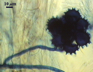
Note variation in height of surface spines in far left photo.
Notes
Immature spores are salmon colored with a slight pink tint (0-10-20-0 to 0-20-60-0 using the INVAM Color Chart). The two layers of the spore wall are near-equivalent thicknesses (1.6-2 µm) before laminae develop in L2, with the outer layer not thickening much further. Sublayers of L2 are so refractive that spores often appear to have a “halo” under a dissecting microscope and on slides. Spores of this species is similar to those of Gi. margarita except for small differences in color (actually a continuum) and greater thickness and refractivity of the L2 layer of the spore wall.
The images below can be uploaded into your browser by clicking on the thumbnail or can be downloaded to your computer by clicking on the link below each image. Please do not use these images for other than personal use without expressed permission from INVAM.
High Resolution Images | |
|---|---|
 |  |
 |  |