Rhizophagus diaphanus
(reference accession WV579B)
Whole Spores | |
|---|---|
|
|
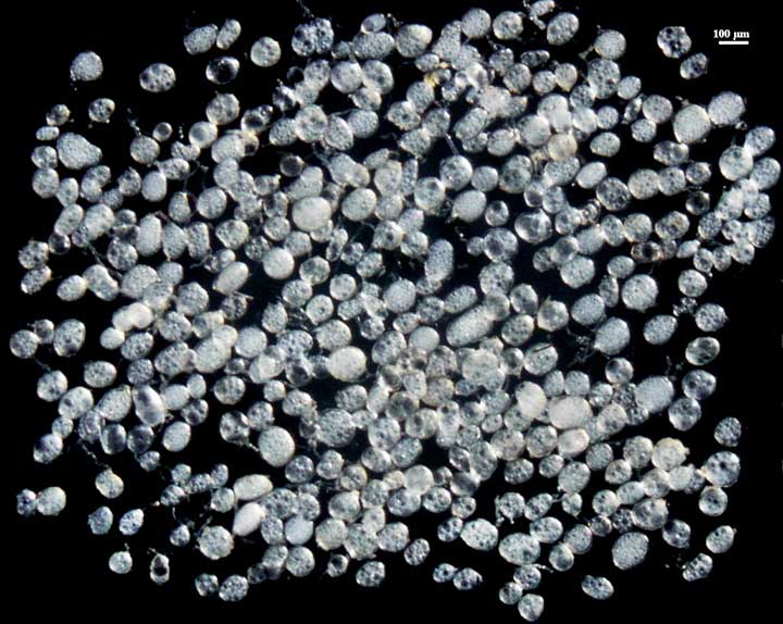
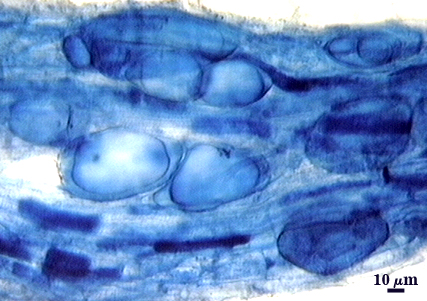
 COLOR: Hyaline to white.
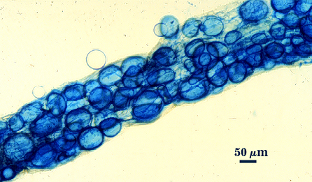
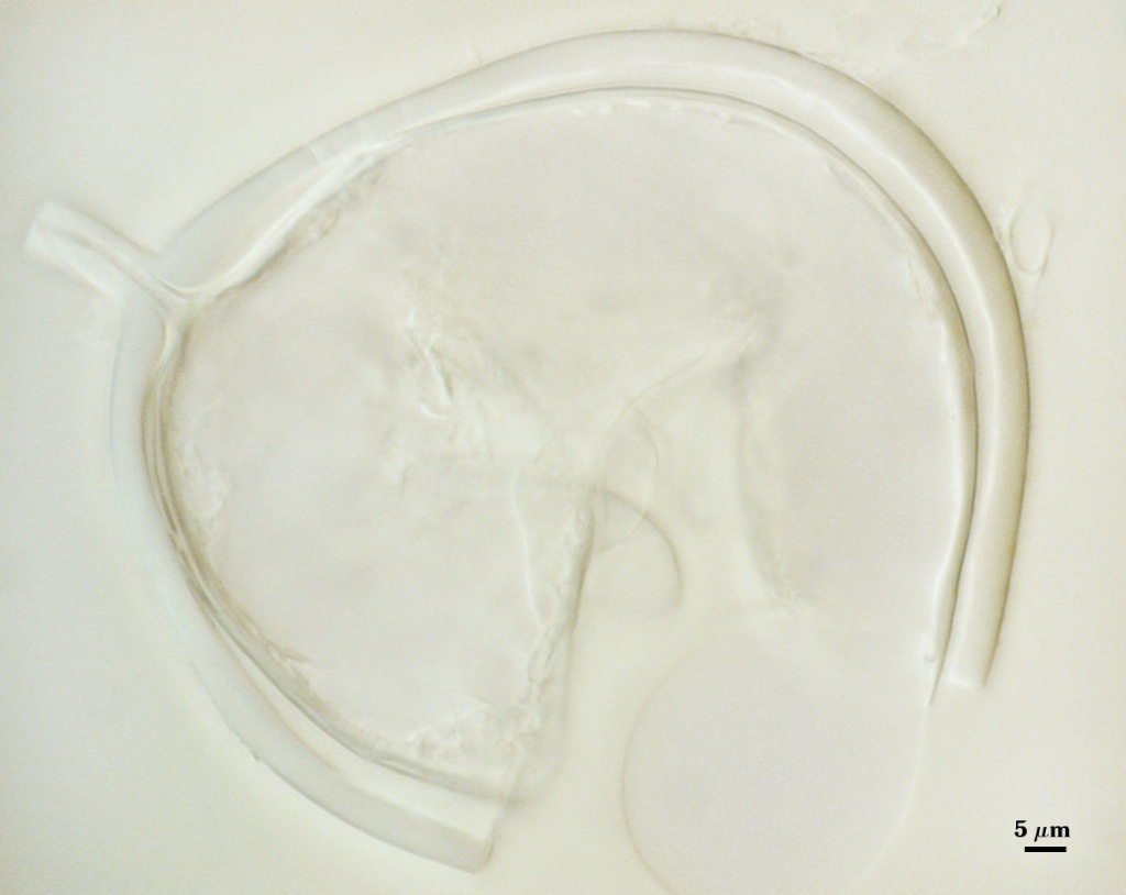
SHAPE: Globose to subglobose, some irregular.
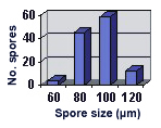
SIZE DISTRIBUTION: 60-120 µm, mean = 93 µm (n = 120).
Subcellular Structure of Spores
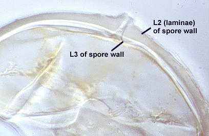
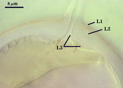
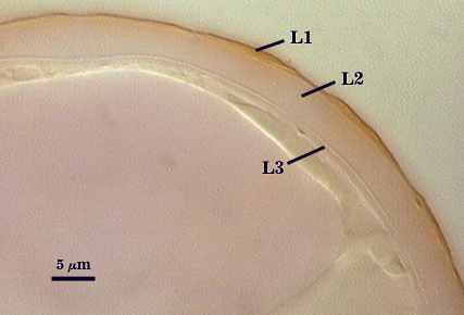
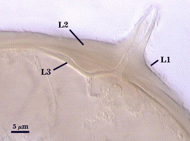
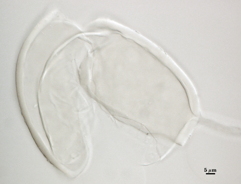
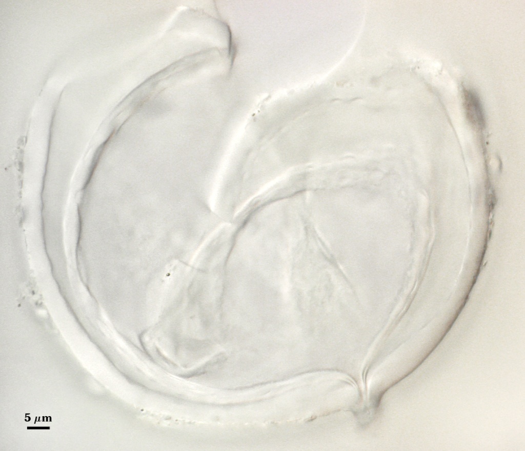
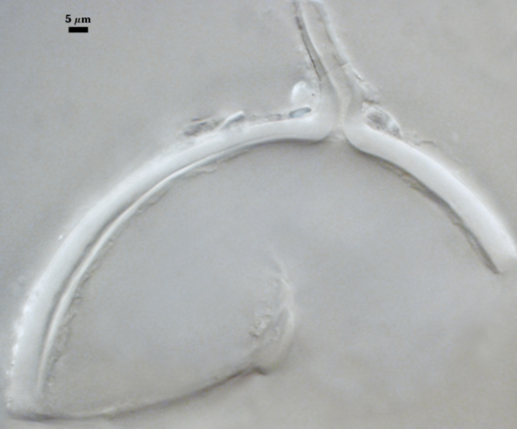
SPORE WALL: Consisting of three layers (L1, L2, and L3) that form consecutively during differentiation of the spore wall.
L1: Very thin mucilagenous layer <0.5 µm) that can stain light pink (0-5-10-0) in melzer's reagent. it is seen rarely populations of mature spores because so thin and light-staining also appears to slough soon after formed (or as a result extraction subsequent washing procedures).
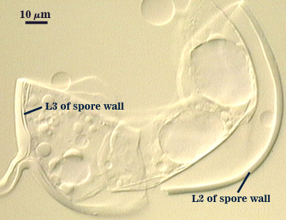
L2: A layer consisting of thin adherent hyaline sublayers (or laminae) that together are very brittle and cause the spore to readily break apart or fragment with only light pressure (it is best to let the slide mountant harden for 30-45 min before applying coverslip to minimize this problem). This layer ranges from 3.2-6.4 µm (mean of 4.9 µm) thick. Some sublayers tend to separate as a group from the others to resemble a separate layer, but this phenotype is rare. Sublayers in the region of subtending hypha can thicken up to 13 µm.
L3: A semi-flexible to flexible layer (depending on thickness), < 0.5-1.6 µm thick (occasionally thickening to 3.0 µm). Even though it has the same phenotype as L2, it is considered a separate layer (instead of a sublayer) because it is detectable in many spores as a separable component. This layer still is part of the spore wall, though, because it is attached to the subtending hyphal wall and thus has a common origin with L1 and L2. It can separate completely from the spore wall in some spores, at which time it resembles a flexible inner wall (synonymous with a “membranous wall” ). It is distinguished from a flexible inner wall by a pore and a protrusion indicating insertion as part of the subtending hyphal wall.
| In PVLG | |
|---|---|
|
|
| In PVLG and Melzer's reagent (1:1 v/v) | |
|---|---|
|
|
Subtending Hypha
SHAPE: Cylindrical to slightly flared (see photos above).
WIDTH: 6.4-11.2 µm (mean = 9.3 µm).
WALL STRUCTURE: Only one layer is evident that is continuous with L2 of the spore wall, 2.2-3.6 µm thick. L1 and L3 may be present, but they are too thin to resolve, even in juvenile spores.
OCCLUSION: L3 of the spore wall often bridges the hyphal pore, resembling a septum.
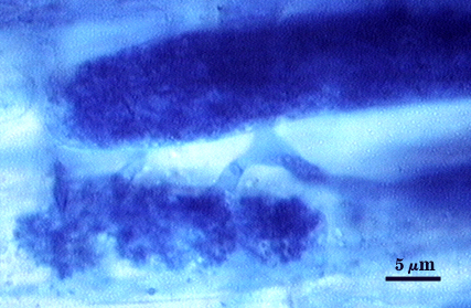
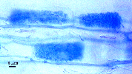
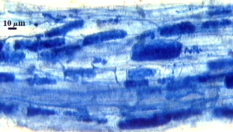
Mycorrhizae
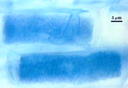
Mycorrhizal components stain darkly in trypan blue. Numerous vesicles often are produced near entry points, and they usually differentiate into spores (as evidenced by a thicker spore wall). Arbuscule production seems to peak earlier than in other fungi in Glomus, with late mycorrhizal colonization consisting almost exclusively of intraradical hyphae and aggregates of spores.
| Arbuscules in cortical cells of corn roots | ||
|---|---|---|
|
|
|
| Arbuscules & hyphae | Sporulation in Maize roots | |
|---|---|---|
|
|
|
Notes
This isolate was recovered from a partially reclaimed coal strip-mine site in northern West Virginia. Paraglomus occultum often is associated with this species in low pH soils (forests, abandoned or reclaimed coal strip-mine sites, etc.). Both are hyaline to white and overlap in size and thus sometimes difficult to distinguish without a little more study. Then, R. diaphanus can be separated by a thin “halo” so that it appears more reflectivein direct lighting under a dissecting microscope. It also readily sinks in water (so that swirling action in a watchglass will cause R. diaphanus spores to accumulate on the bottom, whereas associated P. occultum spores will remain suspended in the water for a short time). In their description of this species, Morton and Walker (1984) fail to mention the presence of an outer mucilagenous layer (L1) and they defined L3 as a “membranous wall” to fit convention at that time. We now know that all three layers are part of one structure (the spore wall), despite differences in appearance (rigid versus folding; adherent versus separating).
Component layers of the spore wall of spores mounted in PVLG for longer than one year become discolored, turning a light tan to dark brown. Therefore, preserved specimens falsely appear to be pigmented.
The spores in roots are highly infective, often more so than soil-borne spores. To increase inoculum potential of isolates of this species, blending of roots is advantageous as it does not appear to cause any reduction in intraradical spore viability.
The images below can be uploaded into your browser by clicking on the thumbnail or can be downloaded to your computer by clicking on the link below each image. Please do not use these images for other than personal use without expressed permission from INVAM.
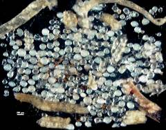
High Resolution Images | |
|---|---|
 |  |
 |  |
 |  |
Reference
- Morton, J. B. and C. Walker. 1984. Glomus diaphanum: A new species in the Endogonaceae common in West Virginia. Mycotaxon 21:431-440.