Diversispora eburnea
(reference accession AZ420A)
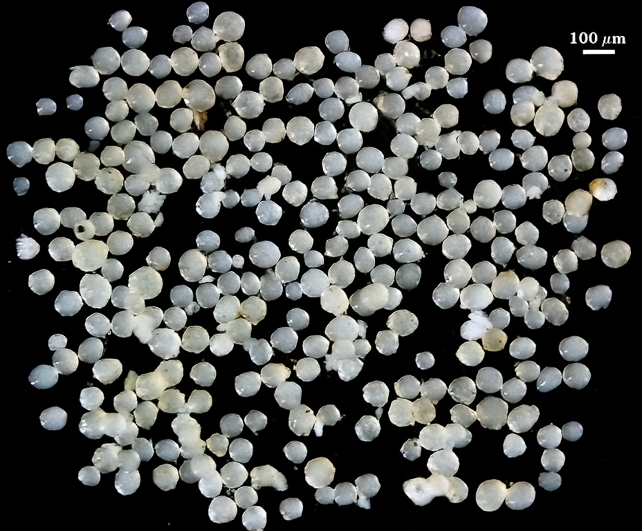
Whole Spores
| Small spores | Large spores |
|---|---|
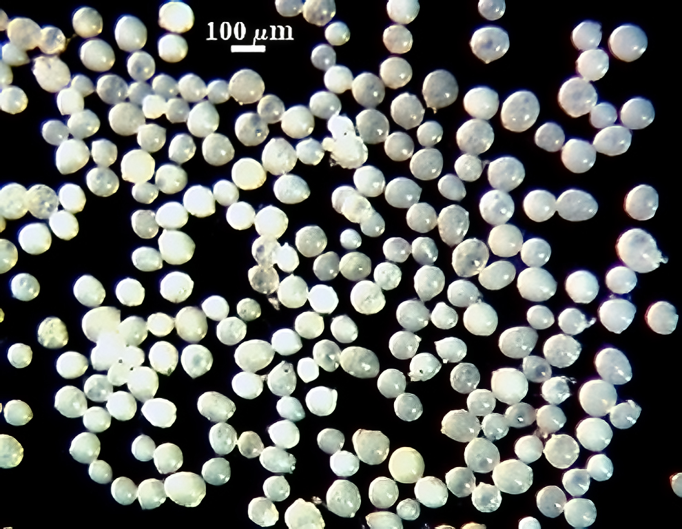 | 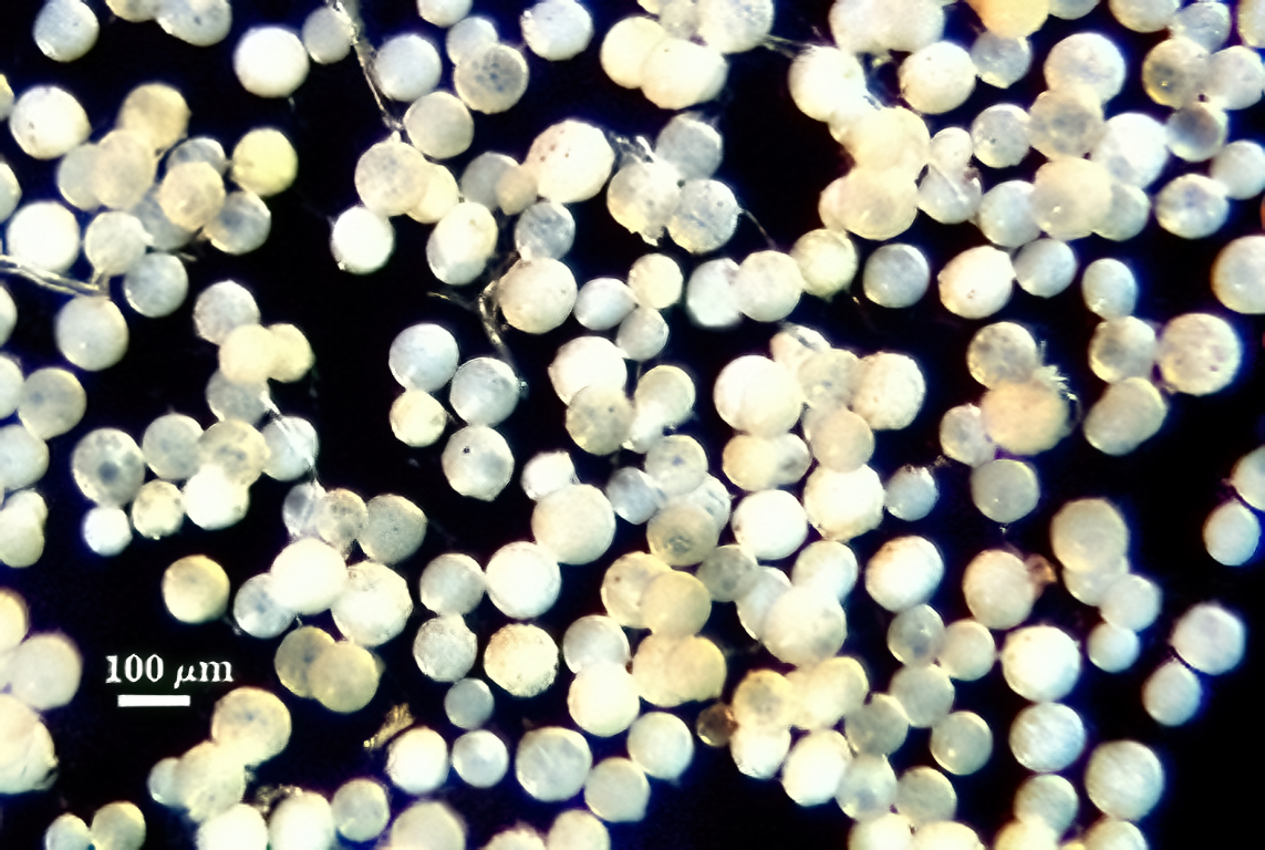 |
 COLOR: Bright white to pale cream (0-0-10-0)
SHAPE: Most often tear-drop shaped to irregular, more rarely globose, subglobose
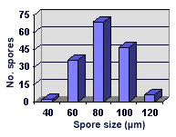
SIZE DISTRIBUTION: 40-60×140-160 µm (globose spores 40-120 µm, mean = 82 µm).
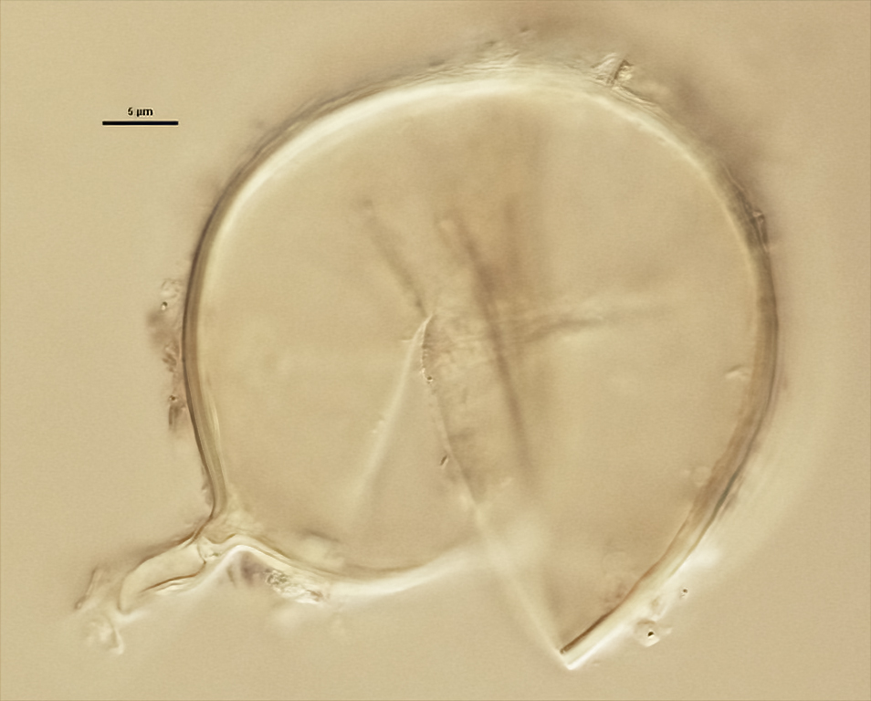
Subcellular Structure of Spores
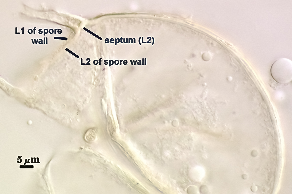
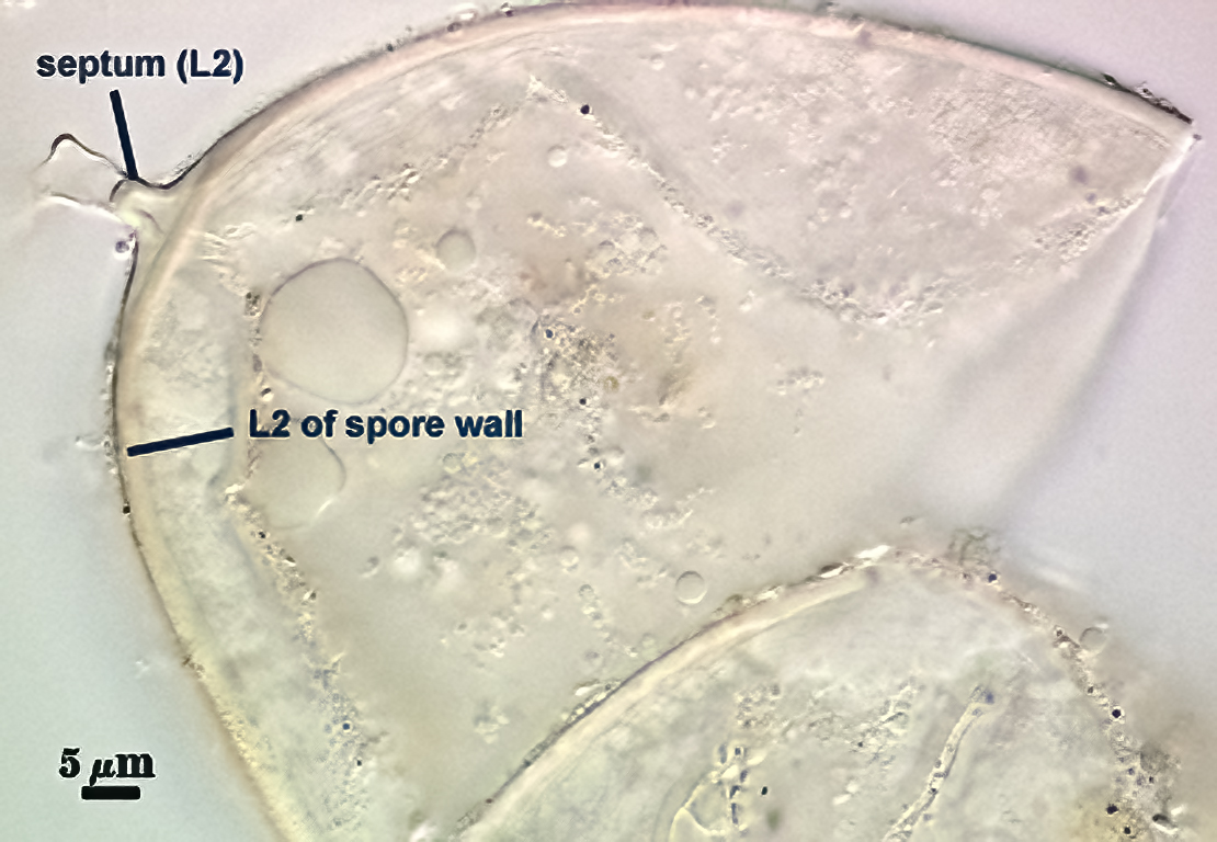
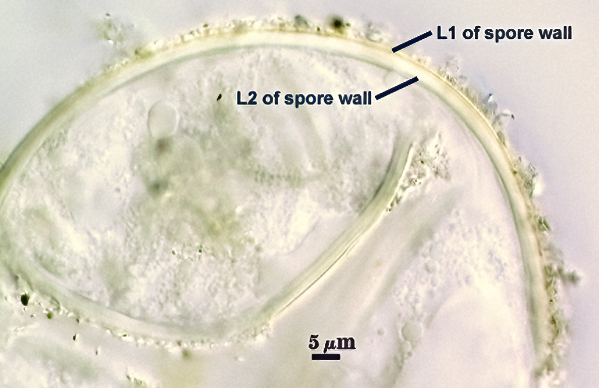
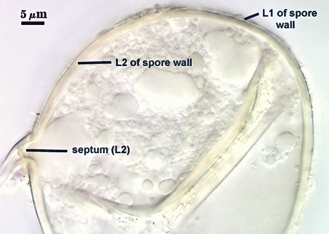
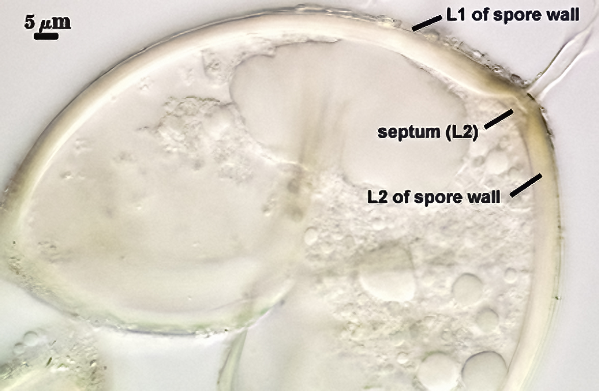
SPORE WALL: Two layers (L1 and L2), L1 usually adherent to L2, even in vigorously crushed spores.
| Spores mounted in PVLG | |||
|---|---|---|---|
 |  |  |  |
| Spores mounted in 1:1 v/v PVLG & Melzer’s reagent | |||
|---|---|---|---|
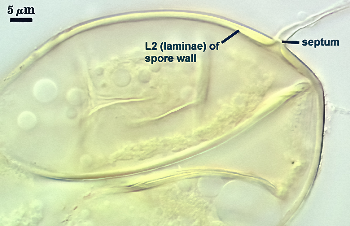 | 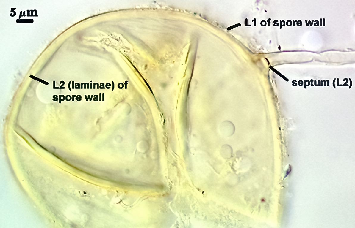 | 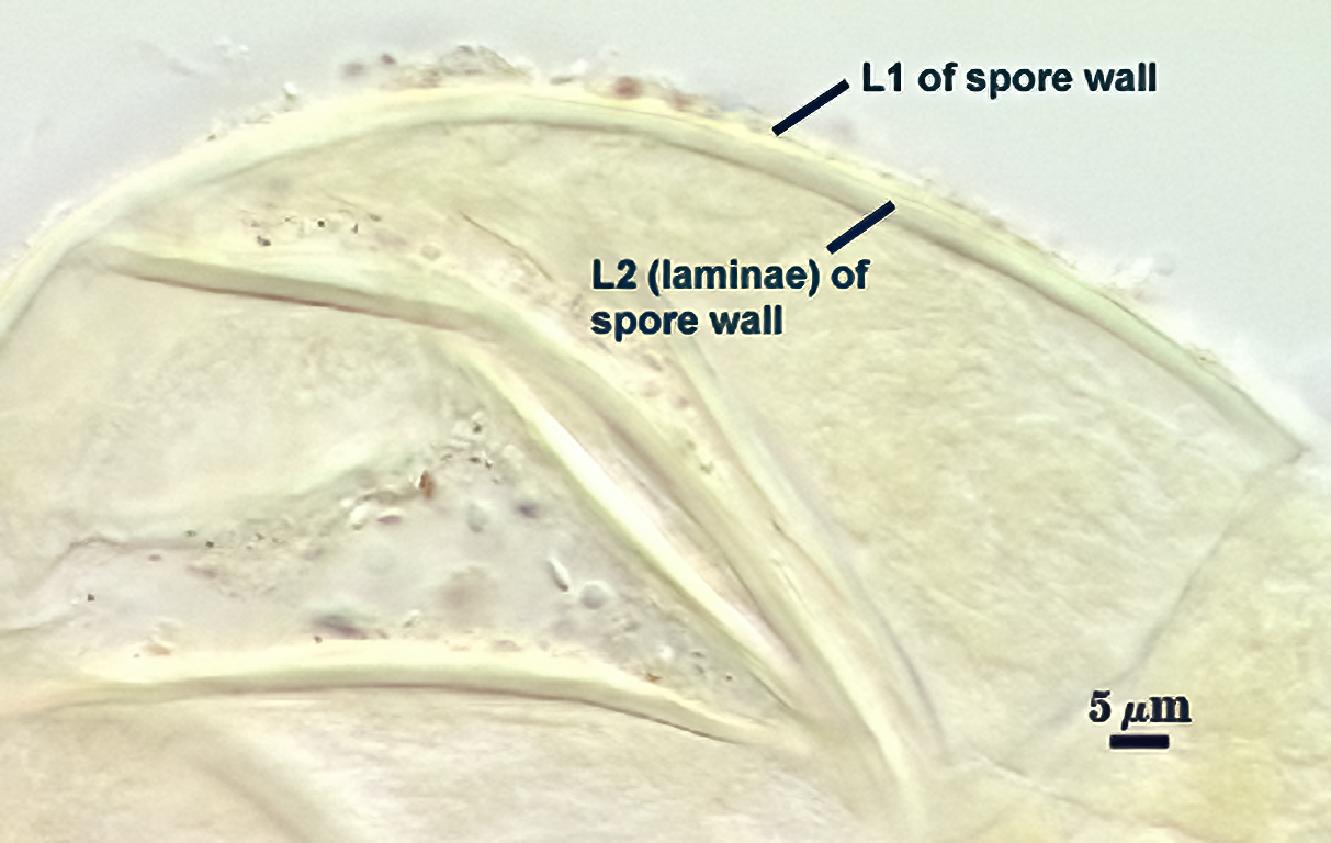 | 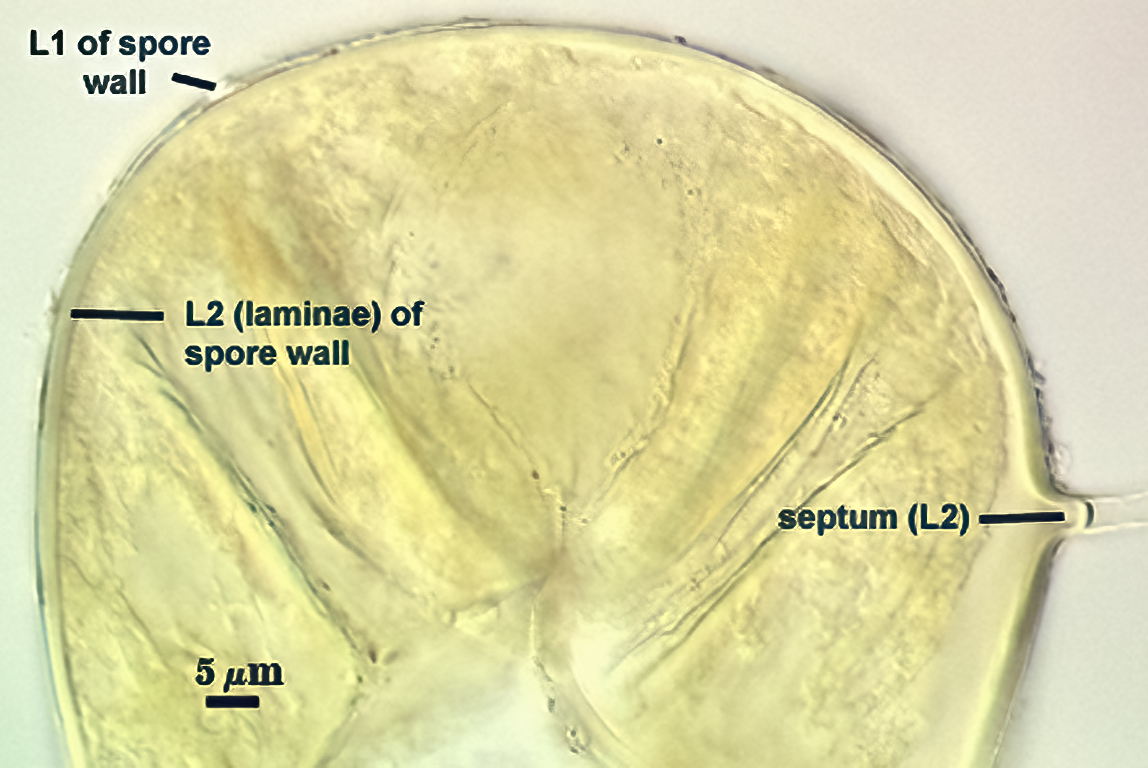 |
L1: Hyaline; 0.5-1.2 µm thick; semi-flexible when separated from L2 (rare), mostly permanent.. Sometimes this layer may be coated with adhering organic debris, especially in the field or in cultures stored for over one year.
L2: Hyaline; consisting of thin hyaline to subhyaline sublayers (or laminae), 1.2-4.0 µm thick (mean = 2.3 µm); semi-flexible to some extent (evident when spores are crushed with a lot of applied pressure).
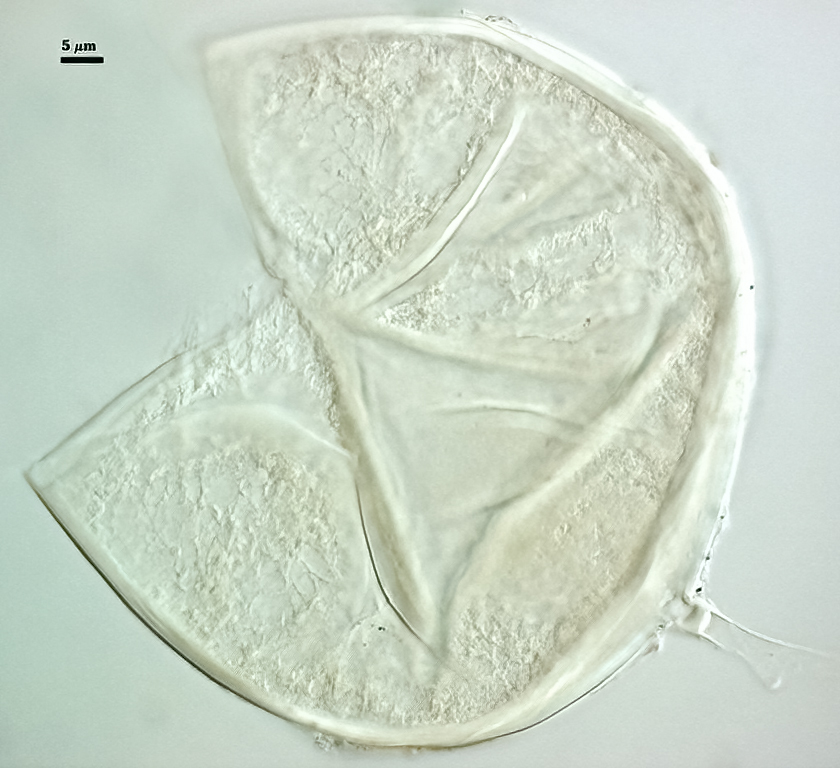
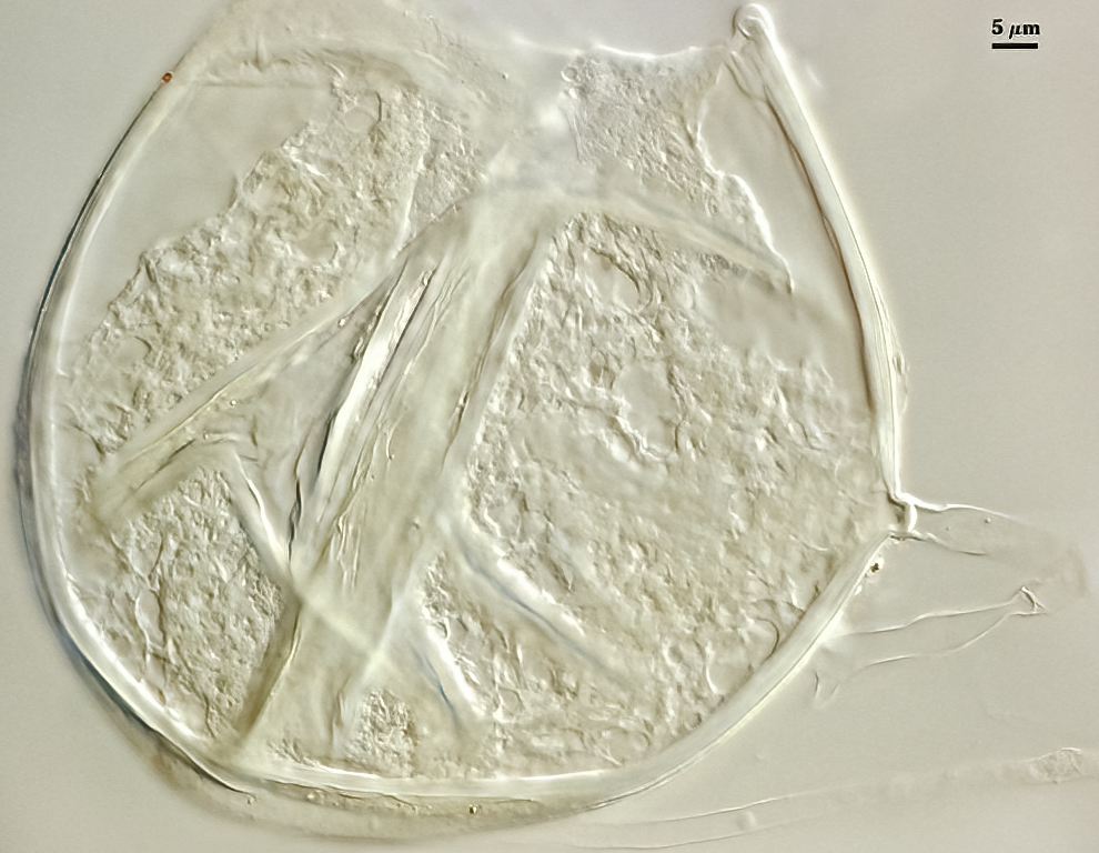
Subtending Hypha
SHAPE: Cylindrical to slightly flared (see photos above and right).
WIDTH: 3.0-6.5 µm (mean = 4.9 µm, n = 40).
WALL STRUCTURE: Two hyaline layers (L1 and L2) that are continuous with the outer two layers of the spore wall. L2 of the spore wall forms a recurved septum, so that only L1 continues further from the spore as the hyphal wall (where it is < 1 µm thick).
OCCLUSION: Usually a hyaline recurved septum, although shape is somewhat variable. Septum is a continuation of L2 of the spore wall and 1.0-2.0 µm thick. It is positioned 1-10 µm in the hypha lumen from point of attachment to the spore (see photos above).
Mycorrhizae
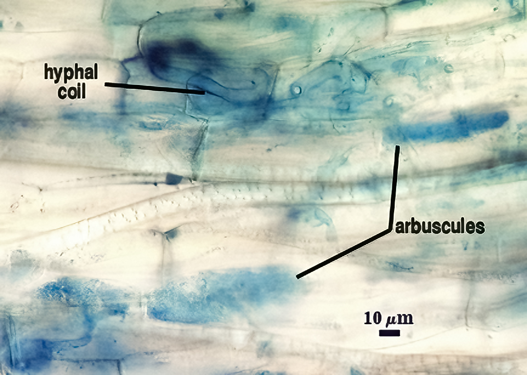
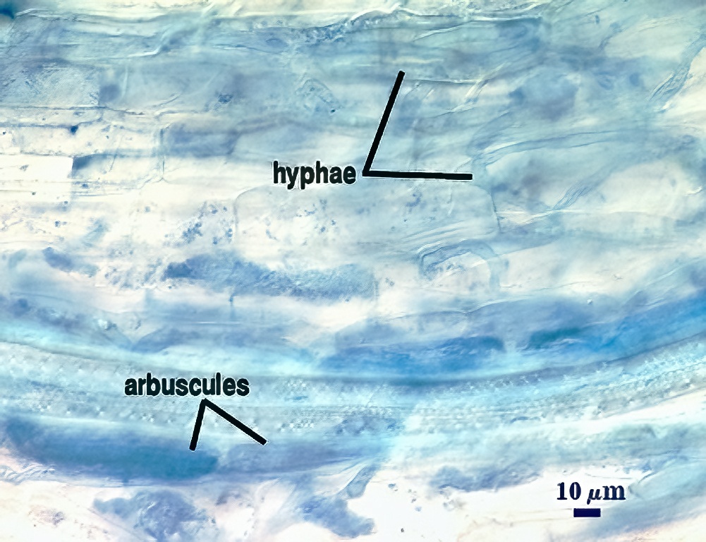
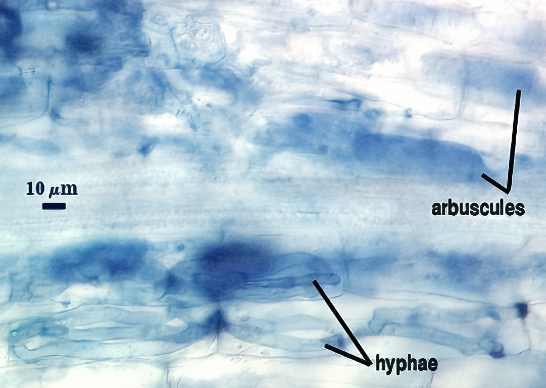
Mycorrhizae in corn roots generally stain faintly in trypan blue, although intensity of the reaction varies from one region of a root to another (or among roots in a sample). Densely branched arbuscules are darkest and become fainter as they senesce. Coiled hphae, many clustered near entry points, are 3-10 µm wide. Straight hyphae are 2-5 µm wide and usually stain only faintly. No vesicle formation was evident, even in 4-month-old cultures (plants already have senesced).
| Arbuscules & intraradical hyphae in corn (digitally enhanced) | |||
|---|---|---|---|
 |  |  |  |
Notes
This species was first cultured from samples in Mexico and was designated in INVAM as “Glomus spurcum-like. With the development of cultures from Arizona, Texas, and West Virginia that grouped distinctly from D. spurca, we realized that it was a distinct species. This lead to a redescription of D. spurca to clearly identify the distinctions between the two species (Kennedy et al., 1999). In soils containing D. eburnea from Arizona and Texas, D. spurca also was present, but we were able to distinguish them sufficiently even at the stereomicroscope level to obtain each in monospecific culture.
The distinguishing features under a stereomicroscope (applied only to freshly collected and healthy spores) is shape (D. eburnea is irregular mostly, D. spurca is globose mostly), appearance of contents (D. eburnea is almost always opaque and bright white, D. spurca is mostly hyaline with transparent centers due to coalescing of lipids). In old/parasitized/senescing field-collected spores or cultured spores stored for 6 months or longer, color and appearance of contents become indistinguishable.
Under a compound microscope, unbroken or very lightly broken spores have a similar spore wall structure. With a little more applied pressure, however, the differences become more evident. L1 often separates from L2 in D. spurca; both layers often remain adherent in D. eburnea. The laminate layer (L2) is much more flexible in D. spurca, so that it produces many folds and gives the appearance of having a thin flexible inner wall, a trait mistakenly reported by Pfeiffer et al. (1996) in the protologue. Even though L2 is slightly flexible in spores of D. eburnea, its not enough to produce similar folds.
The greatest subcellular different between species is in the morphology of transition between spore and subtending hyphal walls and the organization of the occlusion. In D. spurca, a subtending hypha rarely is seen (1 in 50 spores, if lucky) because the hyphal wall is an extension only of L1 of the spore wall, which also is very thin and breaks easily. The laminate layer (L2) thins abruptly and does not extend into the subtending hypha, minimizing any support to keep the hypha attached. In D. eburnea, the subtending hypha is found on most spores because L2 of the spore wall is present to reinforce the hyphal wall in the region of the point of attachment. L2 also forms a substantial septum in the hypha lumen.
The images below can be uploaded into your browser by clicking on the thumbnail or can be downloaded to your computer by clicking on the link below each image. Please do not use these images for other than personal use without expressed permission from INVAM.
High Resolution Images | |
|---|---|
 |  |
 |  |
Links to Gene Sequences in Genbank
Reference
- Pfeiffer, C. M., C. Walker, and H. E. Bloss. 1996. Glomus spurcum: A new endomycorrhizal fungus from Arizona. Mycotaxon 59:373-382.
- Kennedy, J. L., J. C. Stutz, and J. B. Morton. 1999. Glomus eburneum and G. luteum, two previously undescribed species of arbuscular mycorrhizal fungi, with emendation of G. spurcum. Mycologia91:1083-1093.