Glomus fistulosum
(reference accession DN987)
= Glomus claroideum
Whole Spores
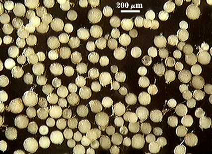
COLOR: Cream (0-0-10-0) to light yellow (0-0-40-0)
SHAPE: Globose to subglobose
SIZE DISTRIBUTION: 80-140 µm, mean = 102 µm
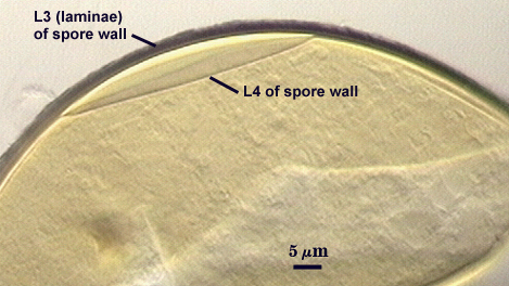
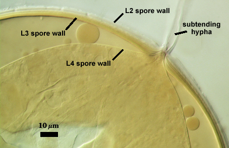
Subcellular Structure of Spores
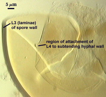
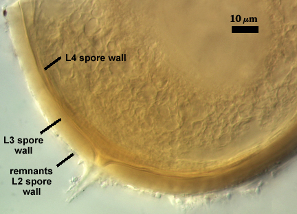
SPORE WALL: Four layers (L1, L2, L3 and L4), only the first two present in juvenile spores and the latter two in mature spores (see sequence for G. claroideum).
| In PVLG | In PVLG & Melzer's reagent | ||
|---|---|---|---|
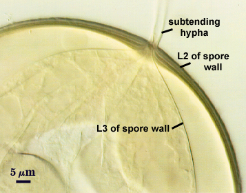 |  |  |  |
L1: Hyaline mucilagenous layer, 0.6-2.1 µm thick in young spores; producing a pink (0-30-20-0) to darker pink (0-40-20-0) reaction in Melzer’s reagent. It often is difficult to distinguish from L2 because both layers are so tightly adherent. This layer appears granular as it decomposes, often sloughing completely in mature spores.
L2: Hyaline, formed in juvenile spores with L1, 0.8-2.0 µm thick, degrading and sloughing concomitant with L1. As this layer degrades, it appears to attract organic debris which can accumulate on the spore surface. No reaction in Melzer’s reagent.
L3: A laminate layer consisting of thin and tightly adherent pale yellow (0-0-20-0) sublayers (= laminations), 2.6-6 µm thick in mature spores.
L4: A thin layer concolorous with the laminate layer, which separates from the remainder of the spore wall in the spore lumen with applied pressure. Formly delimited as a “membranous wall”.
Subtending Hypha
SHAPE: Cylindrical to slightly flared (see photos above)
WIDTH: 5.5-8.4 µm
WALL STRUCTURE: Three layers (L1, L2 and L3) continuous with the layers of the spore wall, 1-3.6 µm thick in the region of attachment, tapering to less than 1.0 µm thick. L1 and L2 usually slough off in mature spores. In many spores, the hyphal wall is so fragile that it breaks from the spore and cannot be seen. L4 does not extend into the hypha beyond the “septum” occluding the spore contents.
OCCLUSION: Either a sublayer of the laminate layer (L3) and L4 or L4 alone bridge the hyphal pore and form what resembles a recurved “septum”.
Germination
A germ tube emerges from the lumen of the subtending hypha.
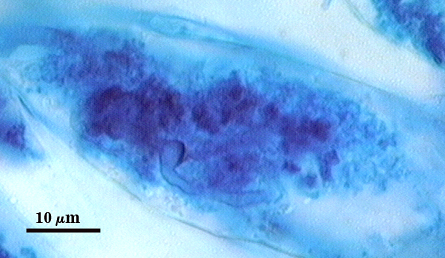
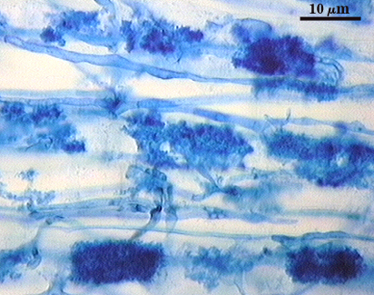
Mycorrhizae
Intraradical hyphae of a mycorrhiza stain darkly in trypan blue. They tend to grow intercellularly between cortical cells, with the hyphal network consisting of numerous “H and h” branches and occasional coils. Some hyphae will have the appearance of projections, but they often are remnants of the trunk hyphae of intracellular arbuscules. Vesicle development is sporadic and patchy, rarely abundant in any particular infection unit. Many mycorrhizae consist of few to no vesicles. Occurrence and abundance of vesicles may be as much environmentally determined as it is genotypic.
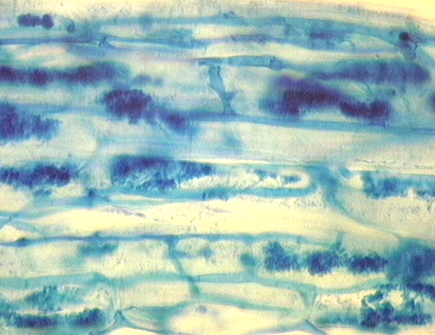
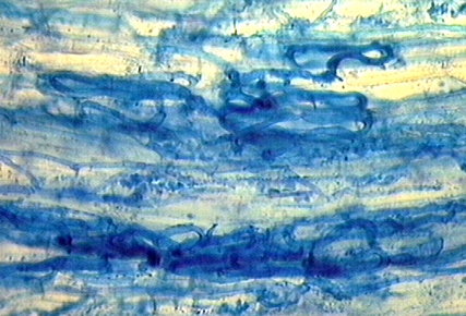
| Arbuscules in corn | |||
|---|---|---|---|
|
|
|
|
Notes
This reference culture is the cultotype contributed by Dr. Skou from a visit in 1987. It has been propagated in four successive cycles and spore morphology has not deviated significantly. All available evidence (morphology of mature spores, developmental pattern) indicated this accession was synonymous with G. claroideum. The complex spore wall structure reported by Skou and Jakobsen (1989) appears to be an artifact of the condition of spores and the way they were crushed when mounted on glass slides. The synonymization of these two species was formalized recently by Walker and Vestberg (1998).
Reference
- Skou, J. P. and I. Jakobsen. 1989. Two new Glomus species from arable land. Mycotaxon 36:273-282.
- Walker, C. and M. Vestberg. 1998. Synonymy amongst the arbuscular mycorrhizal fungi: Glomus claroideum, G. maculosum, G. multisubstensum and G. fistulosum. Annals of Botany 82:601-624.