Funneliformis geosporum
(reference accession CA112)
Whole Spores
| CA112 | MD124 |
|---|---|
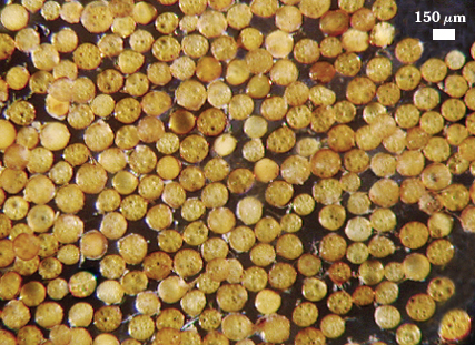 | 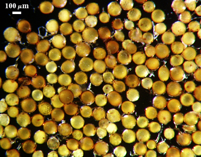 |
 COLOR: Yellow-brown (0-10-40-0) to dark orange-brown (0-30-100-10), the former being more frequent and varying with isolate (see photos above)
SHAPE: Globose to subglobose, some irregular
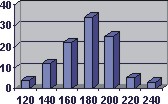
SIZE DISTRIBUTION: 120-240 µm, mean = 176 µm (n = 105)
Subcellular Structure of Spores
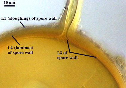
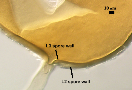
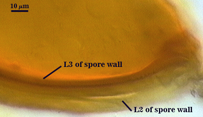
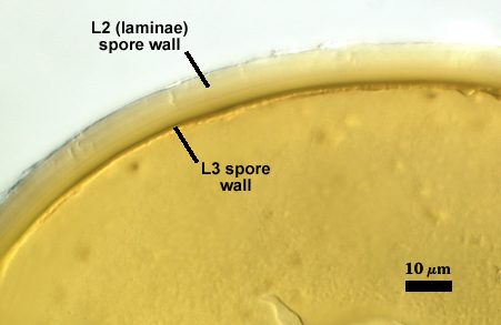
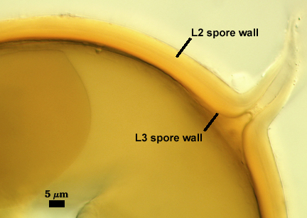
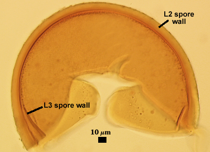
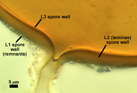
SPORE WALL: Three layers (L1, L2 and L3) that form consecutively as the spore wall differentiates, combined thickness 8-16 µm.
| Spores mounted in PVLG | |||
|---|---|---|---|
 |  |  |  |
| Spores mounted in PVLG and Melzer’s reagent (1:1 v/v) | |||
|---|---|---|---|
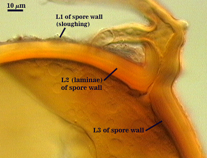 | 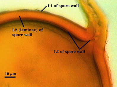 |  |  |
L1: A hyaline layer, < 1 µm thick; not reacting in Melzer’s reagent; degrading and then forming a sloughing granular layer
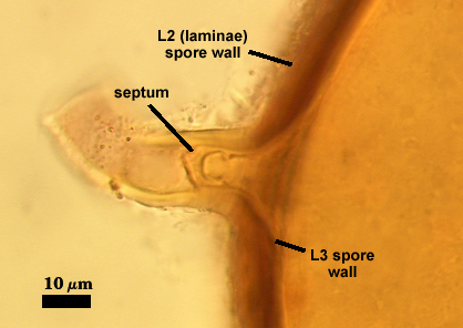
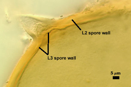
L2: A rigid layer consisting of adherent sublayers (or laminae), yellow-brown to orange-brown in color, often grading from lighter shades on the surface to darker shade toward the inner surface, 6-14 µm thick.
L3: A semi-rigid to rigid layer 1-2.5 µm thick, often adherent to L2, but usually resolved by slightly darker color (yellow to orange-brown), continued growth into the lumen of the subtending hypha to form an unbroken recurved septum. This layer appears to have short stubby warts on the inner surface in some spores; but since this trait is inconsistent, it is not considered a phylogenetically informative structure. We concur with Walker (1982) that this probably is an artifact of microbial activity like that observed in spores of Claroideoglomus claroideum.
Subtending Hypha
SHAPE: Straight to somewhat recurved, often slightly to moderately flared.
WIDTH: 16-32 µm (mean = 24 µm).
WALL STRUCTURE: Only two layers are observed, the outer continuous with L1 and the inner continuous with L3 of the spore wall, together 2.4-4.8 µm in thickness near the spore. The outer layer thins to 1.2-1.6 µm within 50 µm of the spore. Rarely present on the hypha of mature spores. The inner layer is concolorous with L3 of the spore wall, with sublayers adherent.
OCCLUSION: A recurved septum forms 0-30 µm from the point of attachment to the spore (see photos below).
| Smashed Spores | ||
|---|---|---|
 |  |  |
Germination
A germ tube emerges from the lumen of the subtending hypha, originating from the recurved septum. These germ tubes branch extensively after emergence from the hypha.
Notes
Walker (1982) describes Glomus geosporum spores as being most similar to those of G. constrictumbecause of overlap in color, but with enough spores, the distinction is clear. Spores of F. geosporusrarely are darker than a dark orange-brown and are not the red-black to black that dominates in spore populations of S. constrictum. Spores of S. constrictum also do not have a thin inner layer (L3) formed frequently as a component of the spore wall.
Under a stereomicroscope, spores are more likely to be confused with Glomus macrocarpum, which produces spores of similar size and color (our accession MD124 was named G. macrocarpum until LSU sequences placed the isolate in the Funneliformis clade). However, spores do not form a third layer in the spore wall and the spore contents are sealed by thickening of the spore wall (Berch and Fortin, 1983). From available evidence at the time these species were described, both S. constrictumand G. macrocarpum sometimes form spores in loose aggregates, whereas those of F. geosporusare solely ectocarpic and form only singly in the soil. Certainly this is the case of F. geosporus in culture.
Reference
- Berch, S. M. and J. A. Fortin. 1983. Lectotypification of Glomus macrocarpum and proposal of new combinations: Glomus australe, Glomus versiforme, and Glomus tenebrosum (Endogonaceae). Can. J. Bot. 61:2608-2617.
- Walker, C. 1982. Species in the Endogonaceae: a new species (Glomus occultum) and a new combination (Glomus geosporum). Mycotaxon 15:49-61.