Diversispora globifera
(Reference culture NM105)
Whole Spores
| Globifera | ||
|---|---|---|
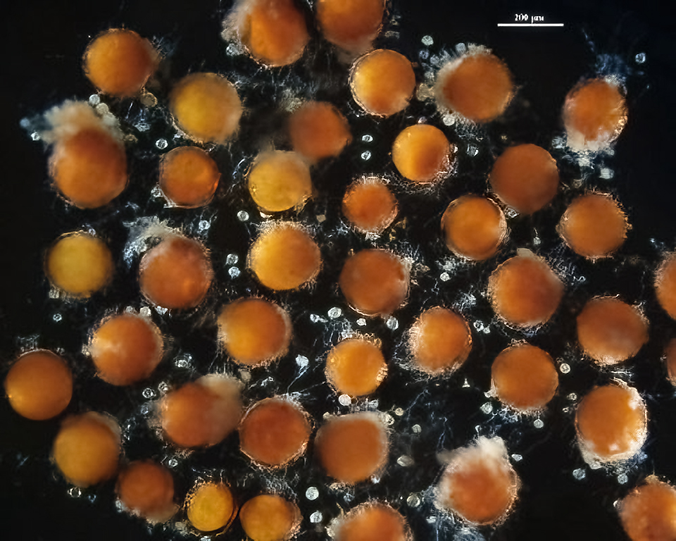 | 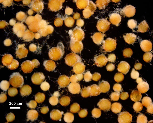 | |
 COLOR: Spores mostly are yellow-brown (0-10-60-0) to orange-brown (0-40-100-10), tortsize.gif (2158 bytes) but they can be dark orange-brown at times (0-20-40-0).
SHAPE: Mostly globose to subglobose; occasionally irregular.
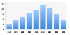
SIZE DISTRIBUTION: 160-320 µm; mean = 249 µm (n = 80)
SPOROCARPS: Many spores occur singly, even when a peridium is present. Less frequently, 2-4 spores will cluster due to intertwining of the peridia amongst the spores.
Subcellular Structure of Spores
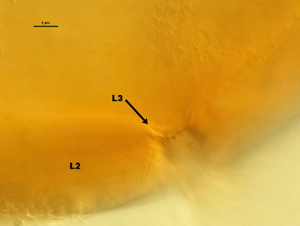
The spore wall (sw) is smooth when no peridium is present. It consists of three layers (L1, L2 and L3) that tend to be tightly adherent most of the time.
| Spore walls | ||
|---|---|---|
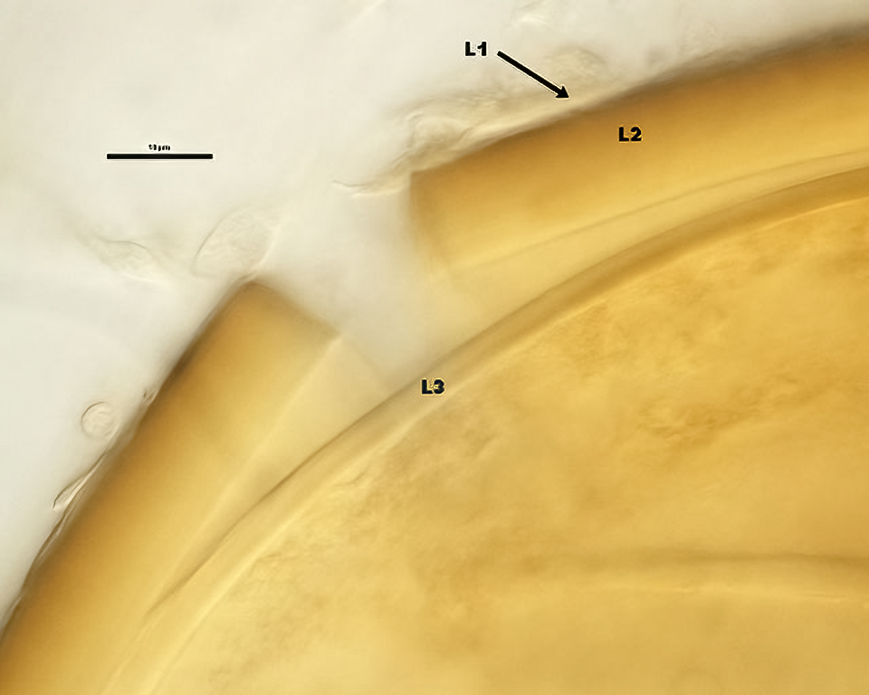 | 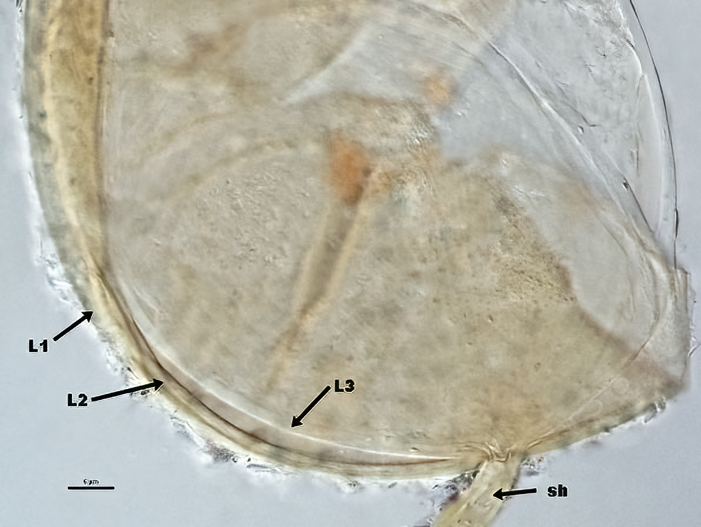 | 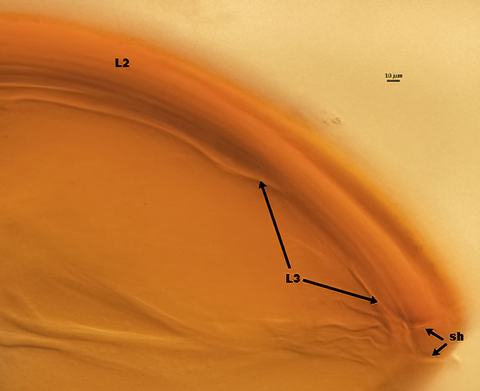 |
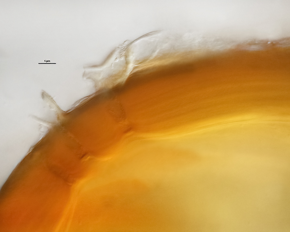
L1: Outermost on spore, of variable thickness (0.5-2 µm) and pale yellow in color. This layer can slough, but it doesn’t degrade nor does it react in Melzer’s reagent. It merges with peridium hyphae when they are present.
L2: Middle layer, which is made up of finely adherent sublayers (or laminae), orange-brown to red-brown in color; 6-30 µm thick, with much of this variation attributed to the plasticity of this layer with applied pressure.
L3: Inner layer, hyaline, also of variable thickness (2-4 µm). Because this layer is transparent, it is difficult to see except when the outer pigmented layers break away. It also appears to be semi-flexible, but does not react in Melzer’s reagent. It spans the lumen of the subtending hypha, functioning like a septum, and thus forms de novo within the spore rather than forming as an extension of the subtending hyphal wall.
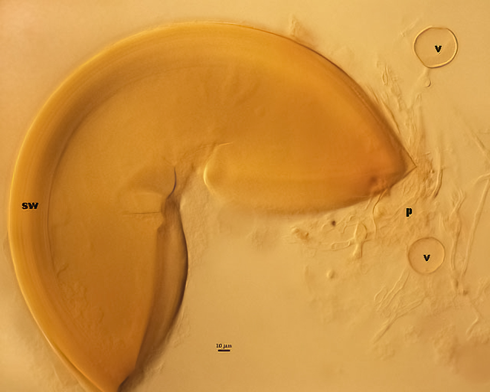
Peridium (p): Originates from branching of the subtending hypha near the spore base when spores are immature and the spore wall is a single thin layer. It consists of loose to tightly interwoven pale yellow to yellow-brown, coenocytic or sparsely septate, thin-walled hyphae, 5-50 µm broad, bearing numerous terminal or intercalary globose to ovoid, pale yellow-brown glomoid swellings (v=vesicles). Vesiculate swellings are 12-75 µm in size, with a wall 1-2 µm thick. Spores without the peridium often dominate in a pot culture of this accession.
Subtending Hypha
| Vesicles | |
|---|---|
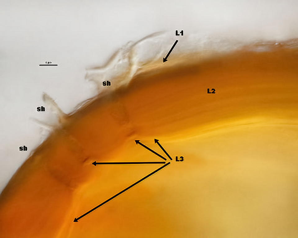 | 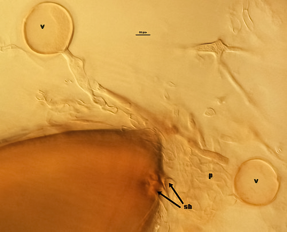 |
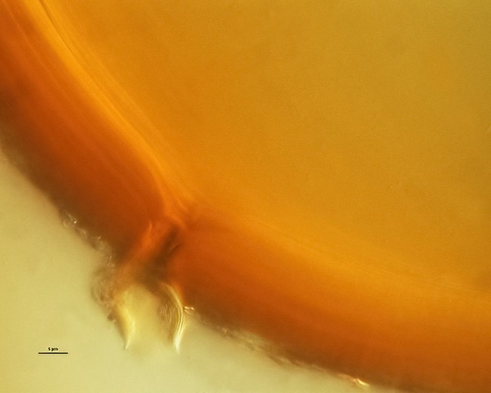
Spores can have only one hypha subtending the base of the spore, but a higher percentage of spores have 2-3 hyphae clustered together. All subtending hyphae tend to be straight or slightly recurved, rarely funnel-shaped; 15-26 µm wide at the spore base with the wall 2-6 µm thick.
Occlusion
The innermost layer of the spore wall (L3) appears to usually bridge the lumen of the subtending hypha, thus functioning much like a septum to compartmentalize spore contents. Koske and Walker (1986) described the subtending hypha as appearing to be “inserted into the spore wall”, but this is not evident in spores from this culture.
Notes
Spores mostly closely resemble those of D. tortuosa. Koske and Walker (1986) describe G. tortuosum spores as being smokey grey-brown color under a stereomicroscope, while those of D. globifera typically are orange-brown to red-brown. This may have been true of their specimens, but the D. tortuosa accession in INVAM are of similar color to the lighter spores of D. globifera. Spores of both species are capable of forming a dense peridium, but that of D. globifera also can form vesiculate swellings.
This accession exhibits the same morphology as the culture descended from type material (FL327) that was deposited by David Sylvia in 1993. LSU sequences of that isolate (which later died) groups with NM105 in a highly supported clade (not yet published).
The images below can be uploaded into your browser by clicking on the thumbnail or can be downloaded to your computer by clicking on the link below each image. Please do not use these images for other than personal use without expressed permission from INVAM.
High Resolution Images | |
|---|---|
| |
| |
 |  |
Links to Gene Sequences in Genbank
Reference
- Koske, R. E. and C. Walker. 1986. Glomus globiferum: a new species of Endogonaceae with a hyphal peridium. Mycotaxon 26:133-142.